 Back
A Tour of the Cell: Structure and Function in Eukaryotic and Prokaryotic Cells
Study Guide - Smart Notes
 Tailored notes based on your materials, expanded with key definitions, examples, and context.
The Cell: Basic Unit of Structure and Function
Overview of Cellular Organization
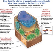
The cell is the fundamental unit of life, providing structure and function for all living organisms. Eukaryotic cells possess internal membranes that compartmentalize their functions, allowing specialized chemical reactions to occur in distinct organelles.
Energy and Material Transformation: Organelles such as mitochondria and chloroplasts convert energy and synthesize essential molecules.
Genetic Information Storage and Transmission: The nucleus stores DNA, which is transcribed and translated by ribosomes to produce proteins.
Interactions with the Environment: The plasma membrane regulates the exchange of substances and signals with the external environment.
Microscopy: Observing Cells
Principles and Types of Microscopy
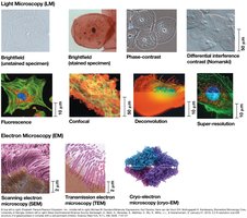
Microscopy enables the observation of cells and their structures, which are too small to be seen with the naked eye. Light microscopes (LM) use visible light and glass lenses to magnify specimens, while electron microscopes (EM) use electron beams for higher resolution.
Magnification: Ratio of image size to actual size.
Resolution: Clarity of the image; minimum distance between two distinguishable points.
Contrast: Difference in brightness between light and dark areas.
Types of Electron Microscopes:
Scanning Electron Microscope (SEM): Produces 3D images of specimen surfaces.
Transmission Electron Microscope (TEM): Reveals internal cell structures.
The Size Range of Cells
Comparative Scale of Biological Structures
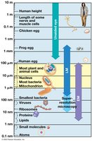
Cells and their components vary greatly in size, from atoms and molecules to entire cells. Most animal and plant cells are 10–100 µm, while prokaryotic cells are 1–5 µm.
Human cells: 10–100 µm
Bacteria: 1–5 µm
Organelles: 0.1–10 µm
Viruses: 0.01–0.1 µm
Prokaryotic vs. Eukaryotic Cells
Basic Features and Classification
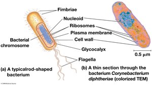
All cells share certain features: plasma membrane, cytosol, chromosomes, and ribosomes. However, prokaryotic and eukaryotic cells differ in complexity and internal organization.
Prokaryotic Cells: No nucleus, DNA in nucleoid, no membrane-bound organelles, smaller size (1–5 µm).
Eukaryotic Cells: DNA in nucleus (double membrane), membrane-bound organelles, larger size (10–100 µm).
Domains: Bacteria and Archaea (prokaryotes); Eukaryotes (protists, fungi, animals, plants).
Eukaryotic Cell Structure
Animal and Plant Cell Organization
Eukaryotic cells contain numerous organelles, each with specialized functions. Internal membranes divide the cell into compartments, facilitating metabolic processes.
Animal Cells: Nucleus, endoplasmic reticulum, Golgi apparatus, mitochondria, lysosomes, peroxisomes, cytoskeleton.
Plant Cells: All animal cell organelles plus chloroplasts, central vacuole, cell wall, plasmodesmata.
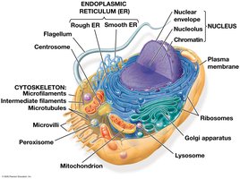
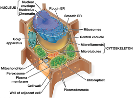
The Nucleus: Information Central
Structure and Function of the Nucleus
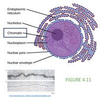
The nucleus is the repository of genetic information, containing most of the cell’s DNA. It is surrounded by a double membrane (nuclear envelope) with nuclear pores for molecular transport.
Chromosomes: DNA molecules associated with proteins, forming chromatin.
Nucleolus: Site of ribosomal RNA (rRNA) synthesis.
Nuclear Envelope: Double lipid bilayer.
Ribosomes: Protein Factories
Structure and Locations of Ribosomes
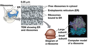
Ribosomes are complexes of rRNA and protein, responsible for protein synthesis. They are found in both prokaryotic (70S) and eukaryotic (80S) cells, either free in the cytosol or bound to the endoplasmic reticulum or nuclear envelope.
Free Ribosomes: Synthesize proteins for use within the cytosol.
Bound Ribosomes: Synthesize proteins for secretion or membrane insertion.
The Plasma Membrane
Structure and Function
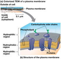
The plasma membrane is a selective barrier composed of a phospholipid bilayer with embedded proteins. It regulates the passage of oxygen, nutrients, and waste, maintaining cellular homeostasis.
Phospholipid Bilayer: Hydrophilic heads face outward, hydrophobic tails inward.
Proteins: Serve as channels, receptors, and enzymes.
The Endomembrane System
Components and Functions
The endomembrane system includes the nuclear envelope, endoplasmic reticulum, Golgi apparatus, lysosomes, vesicles, vacuoles, and plasma membrane. These components are interconnected, regulating protein traffic and metabolic functions.
Nuclear Envelope: Surrounds the nucleus.
Endoplasmic Reticulum (ER): Smooth and rough regions, continuous with nuclear envelope.
Golgi Apparatus: Modifies, sorts, and packages proteins.
Lysosomes: Digestive compartments.
Vesicles/Vacuoles: Storage and transport.
The Endoplasmic Reticulum (ER)
Smooth and Rough ER
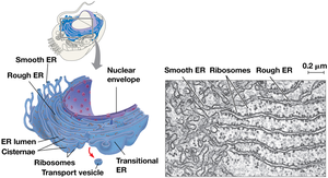
The ER is a biosynthetic factory, accounting for over half the membrane in many eukaryotic cells. The smooth ER lacks ribosomes and is involved in lipid synthesis and detoxification, while the rough ER is studded with ribosomes and synthesizes proteins.
Smooth ER: Synthesizes lipids, metabolizes carbohydrates, detoxifies drugs.
Rough ER: Produces glycoproteins, distributes transport vesicles, membrane factory.
The Golgi Apparatus
Shipping and Receiving Center
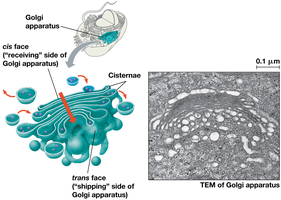
The Golgi apparatus consists of flattened sacs (cisternae) and functions in modifying, storing, and packaging products from the ER. It sorts materials into transport vesicles for delivery to their destinations.
Cis Face: Receiving side.
Trans Face: Shipping side.
Lysosomes: Digestive Compartments
Phagocytosis and Autophagy
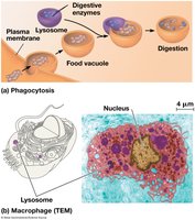
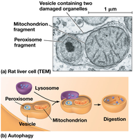
Lysosomes are membranous sacs containing hydrolytic enzymes, primarily found in animal cells. They digest macromolecules, recycle cellular components, and participate in phagocytosis and autophagy.
Phagocytosis: Engulfing and digesting external particles.
Autophagy: Recycling internal organelles and macromolecules.
Vacuoles: Diverse Compartments
Types and Functions of Vacuoles
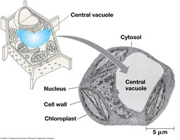
Vacuoles are vesicles derived from the ER and Golgi apparatus. They serve various functions, including storage, water regulation, and maintaining turgor pressure in plant cells.
Food Vacuoles: Formed by phagocytosis.
Contractile Vacuoles: Pump excess water out of cells (in protists).
Central Vacuole: Stores water and nutrients, creates turgor pressure in plant cells.
Mitochondria and Chloroplasts: Energy Conversion
Structure and Function
Mitochondria are the sites of cellular respiration, generating ATP from the breakdown of sugars and fats. Chloroplasts, found in plants and algae, conduct photosynthesis, converting sunlight into chemical energy.
Mitochondria: Smooth outer membrane, folded inner membrane (cristae), matrix, intermembrane space.
Chloroplasts: Thylakoids, stroma, chlorophyll pigment.
Endosymbiont Theory: Evolutionary Origins
Evidence and Implications
The endosymbiont theory proposes that mitochondria and chloroplasts originated from prokaryotic cells engulfed by ancestral eukaryotes. Evidence includes double membranes, circular DNA, ribosomes, and independent reproduction.
Double Membrane: Similar to prokaryotes.
Circular DNA: Resembles bacterial genomes.
Ribosomes: Similar to prokaryotic ribosomes.
The Cytoskeleton: Support and Motility
Components and Functions
The cytoskeleton is a network of fibers that organizes cell structures and activities. It provides structural support, facilitates movement, and anchors organelles.
Microtubules: Thickest, made of tubulin, shape and support, guide organelle movement, chromosome separation.
Microfilaments: Thinnest, made of actin, bear tension, core of microvilli.
Intermediate Filaments: Middle diameter, structural stability.
Extracellular Structures and Cell Junctions
Cell Walls, ECM, and Junctions
Plant cells have cell walls for protection and support, while animal cells have an extracellular matrix (ECM) composed of glycoproteins. Cell junctions facilitate communication and adhesion between cells.
Cell Wall: Primary, secondary, and middle lamella layers.
ECM: Collagen, proteoglycans, fibronectin.
Junctions: Plasmodesmata (plants), tight junctions, desmosomes, gap junctions (animals).
Integration of Cellular Components
Coordination of Functions
Cellular components work together to perform complex functions, such as immune responses, metabolism, and communication. The coordination of organelles, membranes, and cytoskeleton is essential for life.
