 Back
A Tour of the Cell: Structure, Function, and Diversity
Study Guide - Smart Notes
 Tailored notes based on your materials, expanded with key definitions, examples, and context.
Chapter 6: A Tour of the Cell
The Fundamental Units of Life
Cells are the basic units of structure and function in all living organisms. Every organism is composed of one or more cells, which are the simplest collection of matter that can be alive. Despite their diversity, all cells share certain fundamental features.
All organisms are made of cells.
Cells arise from pre-existing cells by division.
Cells can vary greatly in size, shape, and function, but share common structures such as the plasma membrane, cytosol, chromosomes, and ribosomes.
Microscopy and the Study of Cells
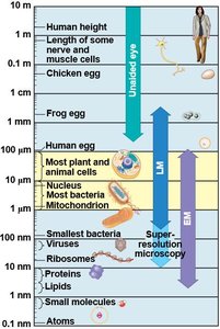
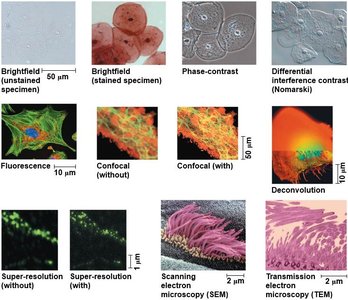
Because cells are typically too small to be seen with the naked eye, microscopes are essential tools for cell biology. Different types of microscopy allow scientists to visualize cells and their components at various levels of detail.
Light Microscopy (LM): Uses visible light and glass lenses to magnify images up to about 1,000 times. Useful for viewing live cells and tissues.
Electron Microscopy (EM): Uses beams of electrons for much higher resolution. Includes:
Scanning Electron Microscopy (SEM): Provides 3D images of cell surfaces.
Transmission Electron Microscopy (TEM): Reveals internal cell structures.
Key Parameters of Microscopy:
Magnification: Ratio of image size to actual size.
Resolution: Clarity of the image; minimum distance between two distinguishable points.
Contrast: Visible differences in brightness between parts of the sample.
Prokaryotic vs. Eukaryotic Cells
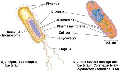
Cells are classified as either prokaryotic or eukaryotic based on their internal structure. This distinction is fundamental to understanding cell biology.
Prokaryotic Cells: Found in Bacteria and Archaea. Lack a nucleus and membrane-bound organelles. DNA is located in the nucleoid region.
Eukaryotic Cells: Found in protists, fungi, animals, and plants. Have a true nucleus enclosed by a nuclear envelope and numerous membrane-bound organelles.
Common Features: All cells have a plasma membrane, cytosol, chromosomes, and ribosomes.
The Plasma Membrane
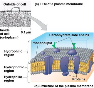
The plasma membrane is a selective barrier that surrounds all cells, controlling the movement of substances in and out. It is primarily composed of a phospholipid bilayer with embedded proteins.
Phospholipid Bilayer: Provides fluidity and forms the basic structure of the membrane.
Proteins: Serve as channels, receptors, and enzymes.
Carbohydrate Side Chains: Involved in cell recognition and signaling.
Internal Organization of Eukaryotic Cells
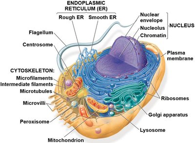
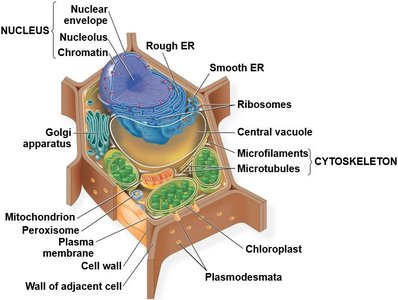
Eukaryotic cells contain numerous organelles, each with specialized functions. These organelles are often surrounded by membranes, allowing compartmentalization of cellular processes.
Nucleus: Contains genetic material (DNA) and is the site of transcription.
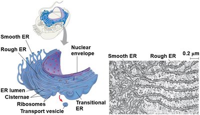
Endoplasmic Reticulum (ER): Involved in protein and lipid synthesis.
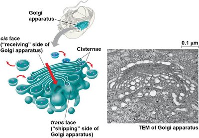
Golgi Apparatus: Modifies, sorts, and packages proteins and lipids.
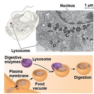
Lysosomes: Digestive organelles containing hydrolytic enzymes.
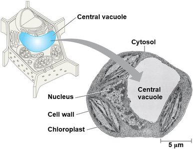
Vacuoles: Storage and maintenance compartments, especially prominent in plant cells.
Mitochondria: Sites of cellular respiration and ATP production.
Chloroplasts: Sites of photosynthesis in plants and algae.
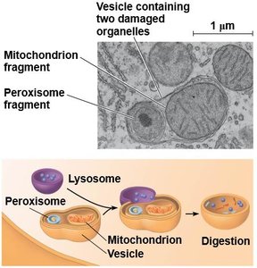
Peroxisomes: Involved in oxidation reactions and detoxification.
The Nucleus and Ribosomes
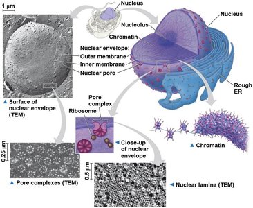
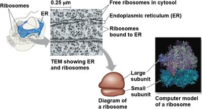
The nucleus is the control center of the cell, housing most of the genetic material. Ribosomes are the sites of protein synthesis, found either free in the cytosol or bound to the ER.
Nuclear Envelope: Double membrane with pores for molecular transport.
Chromatin: DNA-protein complex that condenses to form chromosomes during cell division.
Nucleolus: Site of ribosomal RNA (rRNA) synthesis and ribosome assembly.
The Endomembrane System
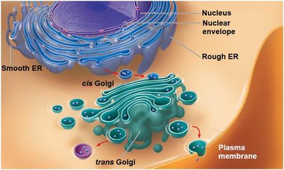
The endomembrane system is a group of membranes and organelles in eukaryotic cells that work together to modify, package, and transport lipids and proteins.
Components: Nuclear envelope, endoplasmic reticulum (ER), Golgi apparatus, lysosomes, vacuoles, and plasma membrane.
Endoplasmic Reticulum (ER):
Smooth ER (SER): Synthesizes lipids, metabolizes carbohydrates, detoxifies drugs, and stores calcium ions.
Rough ER (RER): Studded with ribosomes; synthesizes proteins and membranes.
Golgi Apparatus: Modifies, sorts, and packages proteins and lipids for storage or transport out of the cell.
Lysosomes: Contain hydrolytic enzymes for digestion of macromolecules and recycling of cellular components.
Vacuoles: Storage organelles; central vacuole in plants maintains turgor pressure.
Mitochondria and Chloroplasts: Energy Conversion
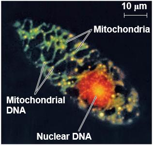
Mitochondria and chloroplasts are the energy-transforming organelles of eukaryotic cells. Mitochondria are the sites of cellular respiration, while chloroplasts carry out photosynthesis in plants and algae.
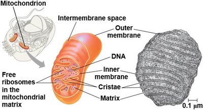
Mitochondria: Double-membraned organelles with their own DNA and ribosomes. The inner membrane is highly folded into cristae, increasing surface area for ATP production.
Chloroplasts: Contain thylakoids (stacked into grana) and stroma. Site of photosynthesis.
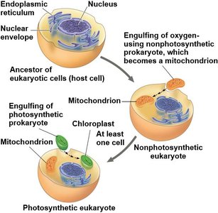
Endosymbiont Theory: Suggests mitochondria and chloroplasts originated as free-living prokaryotes engulfed by ancestral eukaryotic cells.
The Cytoskeleton
The cytoskeleton is a dynamic network of protein fibers that provides structural support, maintains cell shape, and facilitates cell movement and intracellular transport.
Microtubules: Hollow tubes made of tubulin; involved in cell shape, organelle movement, and chromosome separation during cell division.
Microfilaments (Actin Filaments): Thin rods of actin; support cell shape, enable cell movement, and are involved in muscle contraction.
Intermediate Filaments: Fibrous proteins; provide mechanical support and anchor organelles.
Property | Microtubules | Microfilaments | Intermediate Filaments |
|---|---|---|---|
Structure | Hollow tubes | Two intertwined strands of actin | Fibrous proteins coiled into cables |
Diameter | 25 nm with 15-nm lumen | 7 nm | 8-12 nm |
Protein Subunits | Tubulin (α and β) | Actin | Various (e.g., keratins) |
Main Functions | Cell shape, motility, chromosome movement, organelle movement | Cell shape, muscle contraction, cytoplasmic streaming, cell motility, division | Cell shape, anchorage, nuclear lamina formation |
Extracellular Components and Cell Junctions
Cells produce and interact with extracellular materials that help coordinate cellular activities and maintain tissue structure.
Plant Cells: Plasmodesmata are channels that connect plant cells, allowing transport of water, solutes, and signaling molecules.
Animal Cells: Three main types of cell junctions:
Tight Junctions: Prevent leakage of extracellular fluid.
Desmosomes: Anchor cells together into strong sheets.
Gap Junctions: Allow communication and passage of small molecules between cells.
Summary Table: Key Organelles and Their Functions
Organelle | Structure | Function |
|---|---|---|
Nucleus | Double membrane with pores | Houses DNA, site of transcription |
Ribosome | RNA and protein, two subunits | Protein synthesis |
Endoplasmic Reticulum | Network of membranes | Protein and lipid synthesis |
Golgi Apparatus | Stack of cisternae | Modification and sorting of macromolecules |
Lysosome | Membranous sac with enzymes | Digestion and recycling |
Vacuole | Large membrane-bound vesicle | Storage, waste disposal, water balance |
Mitochondrion | Double membrane, cristae | ATP production (cellular respiration) |
Chloroplast | Double membrane, thylakoids | Photosynthesis |
Peroxisome | Single membrane | Oxidation reactions, detoxification |
