 Back
A Tour of the Cell: Structure, Function, and Microscopy
Study Guide - Smart Notes
 Tailored notes based on your materials, expanded with key definitions, examples, and context.
Chapter 4: A Tour of the Cell
Biologists Use Microscopes & Biochemistry to Study Cells
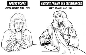
Cell biology relies on microscopy and biochemical techniques to investigate cell structure and function. Early pioneers such as Robert Hooke and Antoni van Leeuwenhoek made foundational discoveries by observing cells and microorganisms.
Microscopy: The use of microscopes to magnify and resolve cellular structures.
Robert Hooke: First recorded person to observe cells and cell walls.
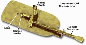
Antoni van Leeuwenhoek: Created his own microscopes and observed 'animalcules' (microorganisms).

Light Microscopy (LM)
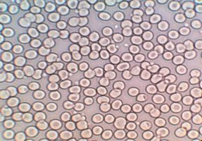
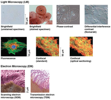
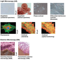
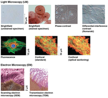
The light microscope uses visible light passed through a specimen and glass lenses to magnify cellular structures. Most cells are too small to be seen by the unaided eye, making microscopy essential for cell biology.
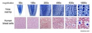
Magnification: Ratio of an object's image size to its real size.
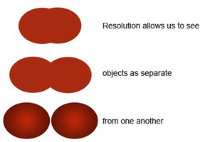
Resolution: Measure of image clarity; minimum distance two points can be distinguished as separate.
Contrast: Difference in brightness between light and dark areas; staining enhances contrast.
Microscopy Parameters and Limitations
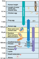
Three key parameters define microscopy: magnification, resolution, and contrast. Light microscopes can magnify up to about 1,000 times, but most subcellular structures are too small to be resolved by LM.
Magnification: Enlarges the image.
Resolution: Determines clarity and ability to distinguish close objects.
Contrast: Enhanced by staining or labeling.
Electron Microscopes
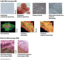
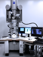
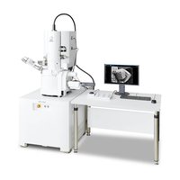
Electron microscopes use beams of electrons, which have much shorter wavelengths than visible light, to study subcellular structures with higher resolution.
Scanning Electron Microscope (SEM): Focuses electron beam on specimen surface, producing 3D images.
Transmission Electron Microscope (TEM): Focuses electron beam through specimen, revealing internal structures.
Recent Advances in Light Microscopy
Modern techniques such as fluorescent labeling and confocal microscopy have improved resolution and allowed visualization of structures as small as 10-20 µm. These advances have led to significant discoveries and Nobel Prizes.
Cell Fractionation
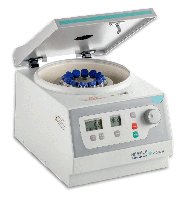
Cell fractionation is a technique where cells are broken up and separated into components by centrifugation. This allows scientists to isolate and study individual organelles.
Cell components separate based on size.
Used to study functions of organelles.
Cell Types and Structure
Prokaryotic vs. Eukaryotic Cells
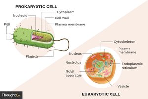
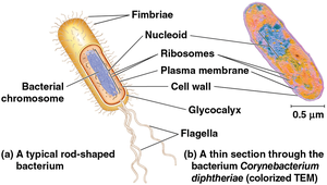
Cells are classified as prokaryotic (Bacteria, Archaea) or eukaryotic (Protists, Fungi, Animals, Plants). All cells share basic features: plasma membrane, cytosol, chromosomes, and ribosomes.
Prokaryotic cells: No nucleus, DNA in nucleoid, no membrane-bound organelles, smaller size (1-5 µm).
Eukaryotic cells: DNA in nucleus, membrane-bound organelles, larger size (10-100 µm).
Plasma Membrane and Surface Area
The plasma membrane is a selective barrier composed of a phospholipid bilayer, allowing passage of oxygen, nutrients, and waste. The ratio of surface area to volume is critical for cell function; smaller cells have a greater ratio, facilitating diffusion.
Nucleus: Genetic Information Center
The nucleus contains most of the cell's DNA, organized into chromosomes. The nuclear envelope separates DNA from cytoplasm, and nuclear pores regulate molecular traffic.
Chromosomes: DNA organized into discrete units.
Chromatin: DNA + proteins, condenses into chromosomes before cell division.
Nucleolus: Site of ribosomal RNA synthesis and ribosome assembly.
Ribosomes: Protein Factories
Ribosomes synthesize proteins using information from DNA. They are composed of ribosomal RNA and protein, and can be found free in the cytosol or bound to the endoplasmic reticulum or nuclear envelope.
Endomembrane System
Components and Functions
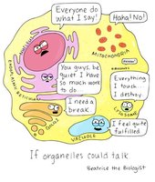
The endomembrane system regulates protein trafficking and metabolic functions. It includes the nuclear envelope, endoplasmic reticulum, Golgi apparatus, lysosomes, vacuoles, and plasma membrane.
Protein synthesis and transport
Metabolism and movement of lipids
Detoxification of poisons
Endoplasmic Reticulum (ER)
The ER is a biosynthetic site, continuous with the nuclear envelope, and accounts for over 50% of the total membrane in eukaryotic cells. It has two regions:
Smooth ER: Lacks ribosomes; synthesizes lipids, metabolizes carbohydrates, detoxifies drugs, stores calcium ions.
Rough ER: Studded with ribosomes; secretes proteins and glycoproteins, distributes proteins via vesicles, membrane factory for the cell.
Golgi Apparatus
The Golgi apparatus consists of flattened sacs (cisternae) and functions as the cell's shipping and receiving center. It modifies ER products, manufactures macromolecules, and sorts/packages materials into vesicles.
Lysosomes
Lysosomes are membranous sacs containing hydrolytic enzymes for digesting macromolecules. They participate in phagocytosis and autophagy, recycling cellular components.
Vacuoles
Vacuoles are large vesicles derived from the ER and Golgi apparatus. They serve various functions:
Food vacuoles: Formed by phagocytosis.
Contractile vacuoles: Pump excess water out of cells in freshwater protists.
Central vacuoles: Found in mature plant cells, store water and ions, and contribute to plant growth.
Energy Organelles
Evolutionary Origins: Endosymbiont Theory
The endosymbiont theory proposes that mitochondria and chloroplasts originated from prokaryotic cells engulfed by ancestral eukaryotes. Evidence includes their double membranes, own DNA, ribosomes, and independent reproduction.
Mitochondria
Mitochondria are the sites of cellular respiration, converting oxygen and organic molecules into ATP. They have a smooth outer membrane and a highly folded inner membrane (cristae), creating compartments for metabolic processes.
Chloroplasts
Chloroplasts are found in plants and algae, and are the site of photosynthesis. They contain chlorophyll, thylakoids (stacked into grana), and stroma (internal fluid with DNA and ribosomes).
Peroxisomes
Peroxisomes are single-membrane organelles containing enzymes that detoxify harmful substances. They convert toxins to hydrogen peroxide (H2O2) and then to water and oxygen.
Cytoskeleton and Cell Structure
Cytoskeleton: Support and Motility
The cytoskeleton is a network of fibers that organizes cell structure and activities, provides mechanical support, and enables cell movement.
Microtubules: Thickest, made of tubulin dimers; shape, support, organelle movement, chromosome separation.
Microfilaments (Actin): Thinnest, bear tension, support cell shape, involved in muscle contraction.
Intermediate filaments: Middle diameter, reinforce cell shape, fix organelles, more permanent.
Extracellular Structures
Cell Walls of Plants
Cell walls are extracellular structures in plants, providing protection, shape, and preventing excessive water uptake. Made of cellulose microfibrils in a matrix of polysaccharides and proteins.
Extracellular Matrix (ECM) of Animal Cells
The ECM is found outside animal cell plasma membranes, composed of glycoproteins (collagen, proteoglycans, fibronectin) and integrins. It binds cells, facilitates communication, and provides structural support.
Cell Junctions
Cell junctions allow neighboring cells to adhere, interact, and communicate:
Plasmodesmata: Channels in plant cell walls for water and solute passage.
Tight junctions: Prevent movement of substances between cells.
Desmosomes: Provide mechanical stability by holding cells together.
Gap junctions: Channels for communication and material passage between animal cells.
Emergent Properties of Cells
Cellular functions arise from the coordinated activity of all cell components. The whole cell exhibits properties greater than the sum of its parts, exemplified by processes such as macrophage-mediated bacterial destruction.
Comparison Table: Prokaryotic vs. Eukaryotic Cells
Feature | Prokaryotic Cells | Eukaryotic Cells |
|---|---|---|
Organisms | Bacteria, Archaea | Protists, Fungi, Animals, Plants |
Nucleus | No (nucleoid region) | Yes (membrane-bound) |
Membrane-bound Organelles | No | Yes |
Size | 1-5 µm | 10-100 µm |
DNA Location | Nucleoid | Nucleus |
Cell Wall | Yes (most) | Plants & fungi only |
Summary Table: Cytoskeleton Components
Component | Diameter | Main Function |
|---|---|---|
Microtubules | Largest | Shape, support, organelle movement, chromosome separation |
Intermediate Filaments | Intermediate | Reinforce shape, fix organelles |
Microfilaments (Actin) | Smallest | Bear tension, support, muscle contraction |
Key Equations
Magnification:
Resolution: where is wavelength and NA is numerical aperture.
