 Back
A Tour of the Cell: Structure, Function, and Diversity
Study Guide - Smart Notes
 Tailored notes based on your materials, expanded with key definitions, examples, and context.
A Tour of the Cell
Introduction to Cell Biology
Cells are the fundamental units of life, and all organisms are composed of one or more cells. Understanding cell structure and function is essential for studying biology, as cells carry out the processes necessary for life. This section provides an overview of cell types, their structural features, and the molecular components that define them.
Cell Size and Microscopy
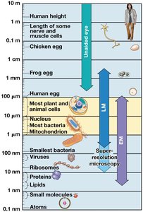
Size Range of Cells
Cells vary greatly in size, from the tiny bacteria to large eukaryotic cells such as eggs. The size of a cell is limited by its metabolic requirements and the ability of its plasma membrane to transport materials efficiently.
Most plant and animal cells are between 10 and 100 micrometers in diameter.
Bacteria are typically 1–5 micrometers in size.
Viruses and ribosomes are much smaller, requiring electron microscopy for visualization.
Surface Area to Volume Ratio
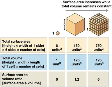
The surface area to volume ratio is a critical factor that limits cell size. As a cell grows, its volume increases faster than its surface area, reducing the efficiency of material exchange across the plasma membrane.
Surface area is important for the exchange of materials (e.g., oxygen, nutrients, waste).
Volume determines the metabolic needs of the cell.
Cells remain small or adopt shapes that increase surface area to maintain efficient exchange.
Total surface area | Total volume | Surface area-to-volume ratio | |
|---|---|---|---|
Single large cell | 6 units2 | 1 unit3 | 6 |
Many small cells | 150 units2 | 125 units3 | 1.2 |
Many tiny cells | 750 units2 | 125 units3 | 6 |
Biological Molecules: Lipids
Overview of Lipids
Lipids are a diverse group of hydrophobic molecules that play key roles in cell structure and function. Unlike other macromolecules, lipids are not true polymers. The main types of biologically important lipids are fats, phospholipids, and steroids.
Fats are constructed from glycerol and fatty acids.
Phospholipids are major components of cell membranes.
Steroids include cholesterol and hormones.
Fats and Fatty Acids
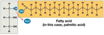
Fats are formed by the combination of glycerol (a three-carbon alcohol) and fatty acids (long hydrocarbon chains with a carboxyl group). Fatty acids can be saturated (no double bonds) or unsaturated (one or more double bonds).
Saturated fatty acids have the maximum number of hydrogen atoms and are typically solid at room temperature.
Unsaturated fatty acids contain double bonds, causing kinks in the chain and are usually liquid at room temperature.
Phospholipids
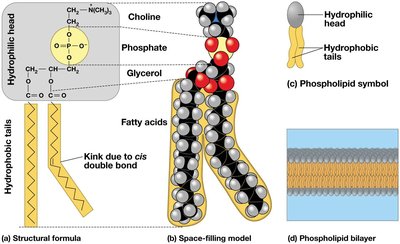
Phospholipids are essential for cell membrane structure. They consist of two hydrophobic fatty acid tails and a hydrophilic phosphate head, allowing them to form bilayers in aqueous environments.
Hydrophilic head: Contains phosphate and is attracted to water.
Hydrophobic tails: Composed of fatty acids, repelled by water.
Phospholipids spontaneously form bilayers, the foundation of all biological membranes.
Steroids
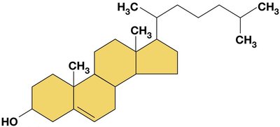
Steroids are lipids with a characteristic structure of four fused carbon rings. Cholesterol is a key steroid in animal cell membranes and serves as a precursor for other steroids, including hormones.
Cholesterol helps maintain membrane fluidity and stability.
Other steroids include sex hormones such as testosterone and estrogen.
Cell Types and Domains of Life
Prokaryotic and Eukaryotic Cells
Cells are classified as prokaryotic or eukaryotic based on the presence or absence of a nucleus and membrane-bound organelles.
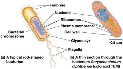
Prokaryotic cells (Bacteria and Archaea): Lack a nucleus and most organelles; DNA is located in the nucleoid region.
Eukaryotic cells (Eukaryotes): Have a true nucleus and numerous membrane-bound organelles.
The Three Domains of Life
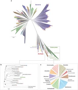
All life is classified into three domains: Bacteria, Archaea, and Eukaryota. Eukaryotes evolved from within Archaea, making them a unique branch of the tree of life. Prokaryotes (Bacteria and Archaea) have dominated Earth's history.
Bacteria: Diverse group of prokaryotes with peptidoglycan cell walls.
Archaea: Prokaryotes with unique membrane lipids and often found in extreme environments.
Eukaryota: Organisms with complex cells containing nuclei and organelles.
Prokaryotic Cell Structure
Cell Envelope and Wall
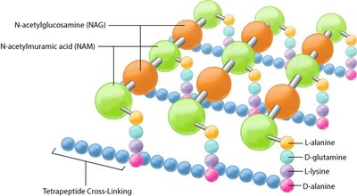
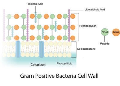
Prokaryotic cells have a cell envelope that may consist of one or two membranes. The cell wall provides structural support and protection. Bacteria build their cell wall out of peptidoglycan, a polymer of sugars and amino acids.
Monoderm (Gram-positive): Single membrane with a thick peptidoglycan layer.
Diderm (Gram-negative): Two membranes with a thin peptidoglycan layer in between.
Unique Features of Archaea
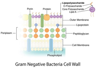
Archaea have unique membrane lipids and cell wall structures that distinguish them from bacteria and eukaryotes. Their membranes often contain ether-linked lipids, and some form monolayers instead of bilayers.
Eukaryotic Cell Structure
General Features
Eukaryotic cells are characterized by compartmentalization, with internal membranes dividing the cell into organelles. This allows for specialized environments and processes within a single cell. Eukaryotic cells are generally much larger than prokaryotic cells.
Nucleus: Contains DNA and is surrounded by a double membrane (nuclear envelope).
Membrane-bound organelles: Include mitochondria, endoplasmic reticulum, Golgi apparatus, and more.
Cytoplasm: Region between the plasma membrane and nucleus.
Many eukaryotes can perform phagocytosis (engulfing particles).
Nucleus and DNA Organization
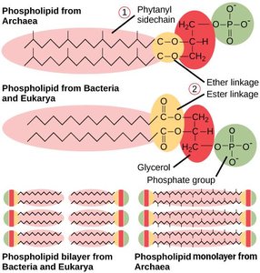
The nucleus is the control center of the eukaryotic cell, containing most of the cell's genetic material. The nuclear envelope is a double membrane that separates the nucleus from the cytoplasm. DNA is organized into chromosomes, and the nucleolus within the nucleus is the site of ribosomal RNA synthesis.
Chromatin: DNA-protein complex that forms chromosomes.
Nucleolus: Site of ribosomal RNA (rRNA) synthesis and ribosome assembly.
Nuclear pores: Regulate entry and exit of molecules from the nucleus.
Ribosomes
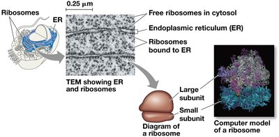
Ribosomes are molecular machines that translate messenger RNA (mRNA) into proteins. They are composed of ribosomal RNA and proteins and can be found free in the cytosol or bound to the endoplasmic reticulum or nuclear envelope in eukaryotes.
Free ribosomes: Synthesize proteins that function in the cytosol.
Bound ribosomes: Synthesize proteins for membranes or export.
Summary Table: Key Differences Between Prokaryotic and Eukaryotic Cells
Feature | Prokaryotic Cells | Eukaryotic Cells |
|---|---|---|
Nucleus | Absent (nucleoid region) | Present (membrane-bound) |
Organelles | Few, not membrane-bound | Many, membrane-bound |
Size | 1–5 μm | 10–100 μm |
Domains | Bacteria, Archaea | Eukaryota |
Cell Wall | Peptidoglycan (Bacteria), various (Archaea) | Cellulose (plants), chitin (fungi), none (animals) |
Additional info: Some humorous or non-standard terms (e.g., "human skin" or "midichlorians") in the provided cartoon are not part of standard cell biology and are omitted from academic explanations.
