 Back
Chapter 3 : Structure, Function, and Microscopy
Study Guide - Smart Notes
 Tailored notes based on your materials, expanded with key definitions, examples, and context.
Exploration of Life Using Microscopes
Types of Microscopy
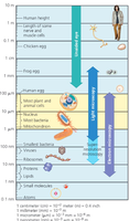
Microscopy is essential for studying cells, which are too small to be seen with the naked eye. Different types of microscopes allow scientists to observe various cellular structures and processes.
Light Microscopy: Uses visible light to illuminate specimens. Suitable for viewing live cells and tissues.
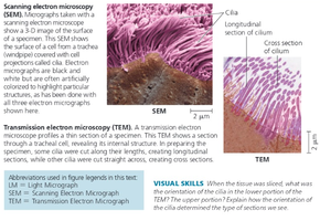
Electron Microscopy: Uses beams of electrons for much higher resolution. Includes:
Scanning Electron Microscopy (SEM): Provides 3D images of cell surfaces.
Transmission Electron Microscopy (TEM): Reveals internal cell structures in thin sections.
Fluorescent Microscopy: Highlights specific molecules using fluorescent tags.
Key concepts in microscopy include:
Magnification: The ratio of an object's image size to its real size.
Resolution: The ability to distinguish two close objects as separate.
Contrast: The difference in brightness between parts of the sample.
Cell Theory and Cell Types
Cell Theory
Cell theory is a fundamental concept in biology:
All living things are composed of cells.
All cells come from pre-existing cells.
Modern additions: DNA is passed during cell division, cells of the same species are structurally and chemically similar, and energy flow occurs within cells.
Prokaryotic vs. Eukaryotic Cells
Cells are classified as either prokaryotic or eukaryotic:
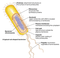
Prokaryotic Cells: Lack a nucleus and membrane-bound organelles. Examples: Bacteria and Archaea.
Eukaryotic Cells: Have a nucleus and various organelles. Examples: Plants, animals, fungi, and protists.
Cell Size, Surface Area, and the Plasma Membrane
Surface Area-to-Volume Ratio
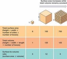
The size of a cell is limited by the surface area-to-volume ratio, which affects the efficiency of material exchange with the environment.
As a cell grows, its volume increases faster than its surface area.
Smaller cells have a higher surface area-to-volume ratio, facilitating efficient exchange of materials.
Small Cube | Medium Cube | Many Small Cubes | |
|---|---|---|---|
Total Surface Area | 6 | 150 | 750 |
Total Volume | 1 | 125 | 125 |
Surface Area-to-Volume Ratio | 6 | 1.2 | 6 |
Plasma Membrane Structure
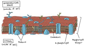
The plasma membrane is a thin, flexible barrier that separates the cell from its environment.
Composed of a phospholipid bilayer with embedded proteins.
Regulates the movement of substances in and out of the cell.
Internal Organization of Eukaryotic Cells
Animal and Plant Cell Structures
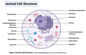
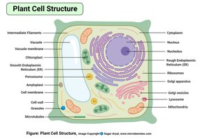
Eukaryotic cells contain various organelles, each with specialized functions.
Nucleus: Stores genetic material and controls cell activities.
Endoplasmic Reticulum (ER): Synthesizes proteins and lipids.
Golgi Apparatus: Modifies, sorts, and packages proteins and lipids.
Lysosomes: Digestive organelles.
Vacuoles: Storage organelles, especially prominent in plant cells.
Mitochondria: Sites of cellular respiration and ATP production.
Chloroplasts: Sites of photosynthesis in plant cells.
Cytoskeleton: Provides structural support and facilitates movement.
The Nucleus and Genetic Control
Nucleus Structure and Function
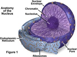
The nucleus is the control center of the cell, containing most of the cell's genetic material.
Chromatin: DNA and proteins, condenses to form chromosomes during cell division.
Nuclear Envelope: Double membrane with nuclear pores for molecular transport.
Nucleolus: Site of ribosomal RNA (rRNA) synthesis and ribosome assembly.
Ribosomes and Protein Synthesis
Structure and Function of Ribosomes
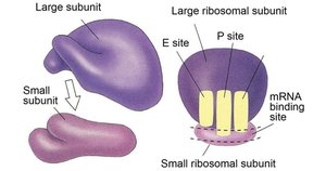
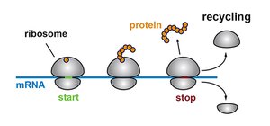
Ribosomes are molecular machines that synthesize proteins by translating messenger RNA (mRNA).
Composed of large and small subunits.
Can be free in the cytosol or bound to the rough ER.
Read mRNA and assemble amino acids into polypeptides.
The Endomembrane System
Components and Functions
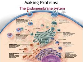
The endomembrane system is a network of membranes involved in the synthesis, modification, and transport of cellular materials.
Endoplasmic Reticulum (ER): Rough ER (with ribosomes) synthesizes proteins; smooth ER synthesizes lipids and detoxifies chemicals.
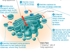
Golgi Apparatus: Modifies and sorts proteins and lipids received from the ER.
Lysosomes: Contain digestive enzymes for breaking down macromolecules.
Vacuoles: Storage and maintenance of cell turgor in plants.
Plasma Membrane: Regulates entry and exit of substances.
Rough vs. Smooth Endoplasmic Reticulum
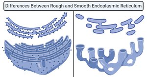
Rough ER: Studded with ribosomes; synthesizes proteins and glycoproteins.
Smooth ER: Lacks ribosomes; synthesizes lipids, detoxifies chemicals, stores calcium ions.
Protein Processing and Transport
Proteins synthesized in the rough ER are packaged into vesicles and transported to the Golgi apparatus for further modification and sorting. 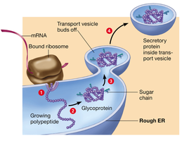
Energy-Related Organelles
Mitochondria
Mitochondria are the powerhouses of the cell, converting chemical energy from food into ATP through cellular respiration.
Double-membrane structure with inner and outer membranes.
Contains its own DNA and ribosomes.
Major site of ATP synthesis.
*Additional info: Mitochondria are believed to have originated from endosymbiotic bacteria.*
Chloroplasts
Chloroplasts are found in plant cells and are the site of photosynthesis.
Double-membrane structure.
Contains stroma (fluid), thylakoids (membranous sacs), and grana (stacks of thylakoids).
Contains its own DNA and ribosomes.
*Additional info: Chloroplasts are also thought to have evolved from endosymbiotic cyanobacteria.*
Cellular Structure and Organization
Cytoskeleton
The cytoskeleton is a network of protein fibers that provides structural support, facilitates cell movement, and organizes organelles.
Microtubules: Hollow tubes of tubulin; involved in cell shape, organelle movement, and chromosome separation.
Intermediate Filaments: Rope-like fibers; provide mechanical strength and anchor organelles.
Microfilaments (Actin Filaments): Thin, flexible fibers; involved in cell movement and muscle contraction.
*Additional info: Motor proteins move along cytoskeletal tracks to transport vesicles and organelles.*
Extracellular Components
Plant Cell Walls
Plant cells are surrounded by a rigid cell wall composed mainly of cellulose.
Primary Cell Wall: Thin and flexible, formed first.
Secondary Cell Wall: Thicker and stronger, formed inside the primary wall.
Middle Lamella: Pectin-rich layer that glues adjacent cells together.
Extracellular Matrix (ECM) in Animal Cells
The ECM is a network of glycoproteins (such as collagen) and other molecules outside animal cells.
Provides structural support and anchorage for cells.
Facilitates cell communication, movement, and growth.
Integrins are membrane proteins that connect the ECM to the cytoskeleton.
*Additional info: The ECM plays a key role in tissue development and repair.*
