 Back
A Tour of the Cell: Structure, Function, and Organization
Study Guide - Smart Notes
 Tailored notes based on your materials, expanded with key definitions, examples, and context.
A Tour of the Cell
Introduction to Cell Biology
Cells are the fundamental units of life, forming the basis of all living organisms. Despite their diversity, all cells share certain structural and functional features that reflect their evolutionary origins.
Cell Theory: All organisms are composed of cells, and all cells arise from pre-existing cells.
Cell Diversity: Cells can vary greatly in size, shape, and function, but all share common characteristics such as a plasma membrane, cytoplasm, genetic material, and ribosomes.
Microscopy and Cell Study
Microscopy Techniques
Microscopes are essential tools for studying cells, which are typically too small to be seen with the naked eye. Different types of microscopes provide varying levels of magnification, resolution, and contrast.
Light Microscopy (LM): Uses visible light and glass lenses to magnify images of specimens. Limited in resolution, making it difficult to study organelles in detail.
Electron Microscopy (EM): Uses electron beams for much higher resolution.
Scanning Electron Microscope (SEM): Provides 3D images of specimen surfaces.
Transmission Electron Microscope (TEM): Used to study internal cell structures.
Key Parameters: Magnification (image size vs. real size), resolution (clarity), and contrast (brightness differences).
Cell Fractionation
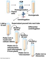
Cell fractionation is a laboratory technique used to separate cellular components for detailed study. It involves homogenizing cells and using differential centrifugation to isolate organelles based on size and density.
Process: Homogenization breaks cells open; centrifugation separates components into pellets (nuclei, mitochondria, microsomes, ribosomes).
Cell Types: Prokaryotic vs. Eukaryotic
Prokaryotic Cells
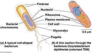
Prokaryotic cells, found in Bacteria and Archaea, lack a nucleus and membrane-bound organelles. Their DNA is located in a region called the nucleoid.
Key Features: Plasma membrane, cytoplasm, ribosomes, nucleoid, cell wall, sometimes flagella and fimbriae.
Example: Corynebacterium diphtheriae is a rod-shaped bacterium with these features.
Eukaryotic Cells
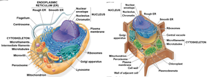
Eukaryotic cells, found in protists, plants, fungi, and animals, have a true nucleus and numerous membrane-bound organelles. They are generally larger and more complex than prokaryotic cells.
Key Features: Nucleus, endoplasmic reticulum, Golgi apparatus, mitochondria, lysosomes, peroxisomes, cytoskeleton, and (in plants) chloroplasts and a central vacuole.
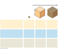
Surface Area-to-Volume Ratio: As cells increase in size, their volume grows faster than their surface area, limiting cell size due to metabolic requirements.
Cell Membranes
Structure and Function of the Plasma Membrane
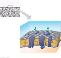
The plasma membrane is a selectively permeable barrier composed of a phospholipid bilayer with embedded proteins. It regulates the passage of substances into and out of the cell, maintaining homeostasis.
Phospholipid Bilayer: Hydrophilic heads face outward; hydrophobic tails face inward.
Proteins: Integral and peripheral proteins serve as channels, receptors, and enzymes.
Carbohydrate Chains: Involved in cell recognition and signaling.
Internal Membranes and Organelles
Nucleus and Ribosomes
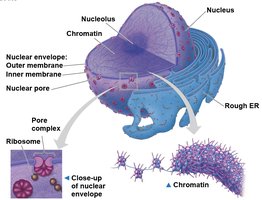
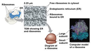
The nucleus houses genetic material and is surrounded by a double membrane (nuclear envelope). Ribosomes, composed of rRNA and protein, are the sites of protein synthesis.
Nucleus: Contains chromatin (DNA + proteins), nucleolus (rRNA synthesis), and nuclear pores for transport.
Ribosomes: Free in cytosol or bound to rough ER; synthesize proteins for various cellular locations.
Endomembrane System
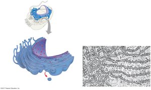
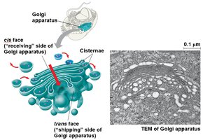
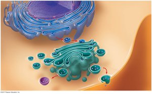
The endomembrane system is a network of membranes involved in protein and lipid synthesis, modification, and transport. It includes the nuclear envelope, endoplasmic reticulum (ER), Golgi apparatus, lysosomes, vacuoles, and plasma membrane.
Endoplasmic Reticulum (ER): Rough ER (with ribosomes) synthesizes proteins; smooth ER synthesizes lipids and detoxifies chemicals.
Golgi Apparatus: Modifies, sorts, and packages proteins and lipids for secretion or delivery to other organelles.
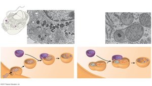
Lysosomes: Contain hydrolytic enzymes for digestion of macromolecules and recycling of cellular components.
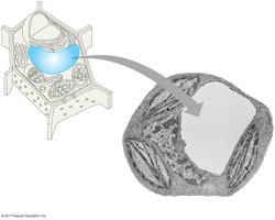
Vacuoles: Storage and maintenance compartments, especially prominent in plant cells (central vacuole).
Energy-Transforming Organelles
Mitochondria and Chloroplasts
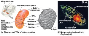
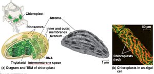
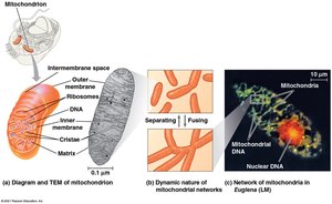
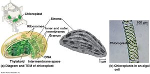
Mitochondria and chloroplasts are organelles that convert energy into forms usable by the cell. Mitochondria perform cellular respiration, while chloroplasts carry out photosynthesis in plants and algae.
Mitochondria: Double-membraned, with inner folds (cristae) and a matrix; contain their own DNA and ribosomes.
Chloroplasts: Contain thylakoids (stacked into grana), stroma, and their own DNA and ribosomes.
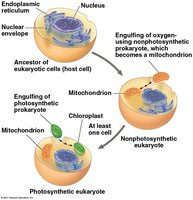
Endosymbiont Theory: Proposes that mitochondria and chloroplasts originated as free-living prokaryotes engulfed by ancestral eukaryotic cells.
Peroxisomes
Peroxisomes are small organelles that detoxify harmful substances and break down fatty acids. They produce hydrogen peroxide as a byproduct, which is then converted to water.
The Cytoskeleton
Structure and Function
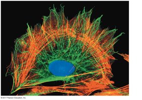
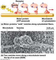
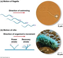
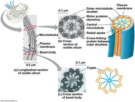
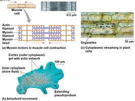
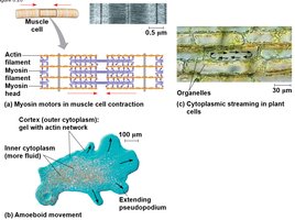
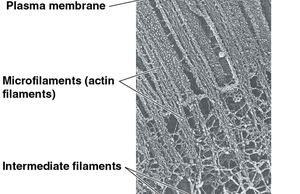
The cytoskeleton is a dynamic network of protein fibers that provides structural support, facilitates cell movement, and organizes organelles within the cell.
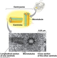
Microtubules: Hollow rods made of tubulin; involved in cell shape, organelle movement, and chromosome separation during cell division.
Microfilaments (Actin Filaments): Solid rods of actin; support cell shape, enable cell movement, and form the core of microvilli.
Intermediate Filaments: Fibrous proteins; provide mechanical support and anchor organelles.
Extracellular Structures and Cell Junctions
Plant Cell Walls
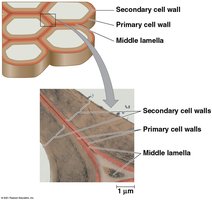
Plant cells are surrounded by a rigid cell wall composed of cellulose. The wall provides structural support, protection, and prevents excessive water uptake.
Layers: Primary cell wall, middle lamella (pectins), and sometimes a secondary cell wall.
Extracellular Matrix (ECM) in Animal Cells
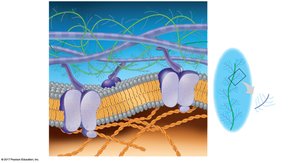
Animal cells lack cell walls but are embedded in an extracellular matrix composed of glycoproteins (collagen, proteoglycans, fibronectin) that provide structural and biochemical support.
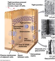
Cell Junctions
Cells in multicellular organisms are connected by specialized junctions that facilitate communication and adhesion.
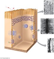
Tight Junctions: Seal neighboring cells to prevent leakage of extracellular fluid.
Desmosomes: Anchor cells together into strong sheets.
Gap Junctions: Provide cytoplasmic channels for communication between adjacent cells.
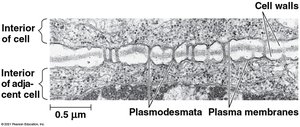
Plasmodesmata (plants): Channels that traverse cell walls, allowing transport and communication between plant cells.
Summary Table: Prokaryotic vs. Eukaryotic Cells
Feature | Prokaryotic Cells | Eukaryotic Cells |
|---|---|---|
Nucleus | Absent (nucleoid region) | Present (membrane-bound) |
Membrane-bound Organelles | Absent | Present |
Size | Small (1–10 μm) | Larger (10–100 μm) |
Examples | Bacteria, Archaea | Protists, Plants, Fungi, Animals |
