Back
A Tour of the Cell: Structure and Function of Prokaryotic and Eukaryotic Cells
Study Guide - Smart Notes
 Tailored notes based on your materials, expanded with key definitions, examples, and context.
The Fundamental Units of Life
Introduction to Cells
Cells are the basic units of life, forming the simplest collection of matter that can be considered alive. All living organisms are composed of cells, which share common features but can differ greatly in structure and function. Cells arise from the division of pre-existing cells, maintaining continuity of life.
Types of Cells: Prokaryotic and Eukaryotic
Prokaryotic Cells
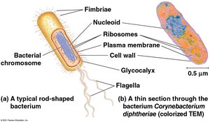
Prokaryotic cells are found in the domains Bacteria and Archaea. They are generally smaller and simpler than eukaryotic cells, lacking a nucleus and membrane-bound organelles.
No nucleus: Genetic material is located in a region called the nucleoid.
No membrane-bound organelles: Cellular functions occur in the cytoplasm.
Cell wall: Provides structural support and protection.
Plasma membrane: Encloses the cell, controlling the movement of substances in and out.
Ribosomes: Sites of protein synthesis.
Other structures: May include fimbriae, flagella, and a glycocalyx for attachment and movement.
Eukaryotic Cells
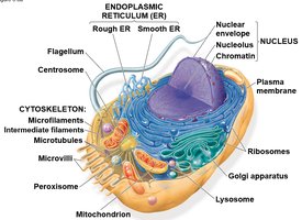
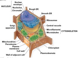
Eukaryotic cells are found in protists, plants, fungi, and animals. They are typically larger and more complex than prokaryotic cells, with compartmentalization of functions into membrane-bound organelles.
Nucleus: Contains the cell's genetic material, enclosed by a double membrane.
Membrane-bound organelles: Specialized structures such as mitochondria, endoplasmic reticulum, Golgi apparatus, and lysosomes.
Cytoplasm: Region between the plasma membrane and the nucleus.
Cell Membranes and Surface Area
Structure of the Plasma Membrane
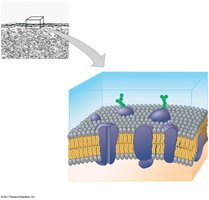
The plasma membrane is a selectively permeable barrier composed of a phospholipid bilayer with embedded proteins. It regulates the passage of oxygen, nutrients, and waste products to maintain cellular homeostasis.
Phospholipid bilayer: Hydrophilic heads face outward, hydrophobic tails face inward.
Proteins: Serve as channels, receptors, and enzymes.
Carbohydrate side chains: Involved in cell recognition and signaling.
Surface Area to Volume Ratio
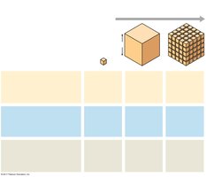
The surface area to volume ratio is critical for cell function. As a cell increases in size, its volume grows faster than its surface area, limiting the rate of material exchange with the environment. This sets an upper limit on cell size.
Small cells: Have a higher surface area to volume ratio, facilitating efficient exchange of materials.
Large cells: May develop adaptations such as folds or projections to increase surface area.
The Nucleus and Ribosomes
Nucleus
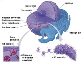
The nucleus is the control center of the eukaryotic cell, housing most of the cell's DNA. It is surrounded by a double membrane called the nuclear envelope, which contains nuclear pores for the exchange of materials with the cytoplasm.
Chromatin: DNA associated with proteins, organized into chromosomes during cell division.
Nucleolus: Site of ribosomal RNA (rRNA) synthesis and ribosome assembly.
Ribosomes
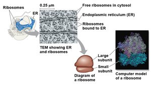
Ribosomes are molecular machines composed of rRNA and proteins. They are responsible for protein synthesis and can be found free in the cytosol or bound to the endoplasmic reticulum (ER).
Free ribosomes: Synthesize proteins that function in the cytosol.
Bound ribosomes: Synthesize proteins destined for membranes, organelles, or secretion.
The Endomembrane System
Components and Functions
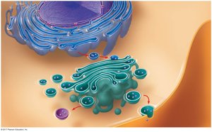
The endomembrane system is a network of membranes within eukaryotic cells that regulates protein traffic and performs metabolic functions. It includes the nuclear envelope, endoplasmic reticulum, Golgi apparatus, lysosomes, vacuoles, and the plasma membrane.
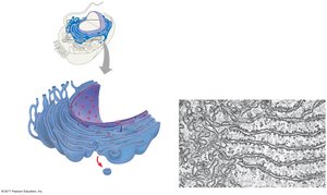
Endoplasmic reticulum (ER): Smooth ER synthesizes lipids and detoxifies chemicals; rough ER is involved in protein synthesis and modification.
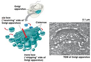
Golgi apparatus: Modifies, sorts, and packages proteins and lipids for storage or transport.
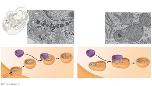
Lysosomes: Contain hydrolytic enzymes for digestion of macromolecules.
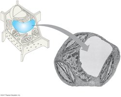
Vacuoles: Storage and maintenance compartments, especially prominent in plant cells.
Energy-Transforming Organelles
Mitochondria
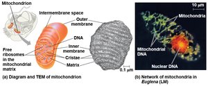
Mitochondria are the sites of cellular respiration, a process that converts chemical energy from food into ATP, the cell's energy currency. They are bounded by a double membrane and contain their own DNA and ribosomes.
Inner membrane: Folded into cristae to increase surface area for ATP production.
Matrix: Contains enzymes, mitochondrial DNA, and ribosomes.
Chloroplasts
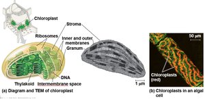
Chloroplasts are found in plants and algae and are the sites of photosynthesis. They convert solar energy into chemical energy stored in glucose. Like mitochondria, they have a double membrane and their own DNA.
Thylakoids: Membranous sacs stacked into grana, where light-dependent reactions occur.
Stroma: Fluid surrounding the thylakoids, site of the Calvin cycle.
The Cytoskeleton
Structure and Function
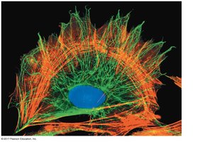
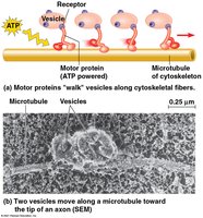
The cytoskeleton is a dynamic network of protein fibers that provides structural support, maintains cell shape, and facilitates cell movement and intracellular transport.
Microfilaments: Composed of actin, involved in cell movement and shape changes.
Intermediate filaments: Provide mechanical support for the cell.
Microtubules: Hollow tubes that serve as tracks for organelle movement and are involved in cell division.
Extracellular Components and Cell Junctions
Plant Cell Walls
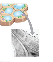
Plant cells are surrounded by a rigid cell wall composed mainly of cellulose. The cell wall provides structural support, protection, and regulates water intake.
Extracellular Matrix (ECM) of Animal Cells
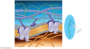
The ECM is a complex network of glycoproteins, such as collagen, proteoglycans, and fibronectin, that provides structural and biochemical support to animal cells. It also plays a role in cell signaling and adhesion.
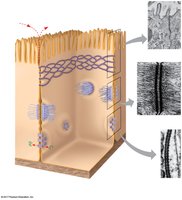
Cell Junctions
Animal cells are connected by specialized junctions that facilitate communication and adhesion:
Tight junctions: Prevent leakage of extracellular fluid between cells.
Desmosomes: Anchor cells together, providing mechanical stability.
Gap junctions: Allow the passage of ions and small molecules between adjacent cells for communication.