 Back
A Tour of the Cell: Structure, Function, and Organization
Study Guide - Smart Notes
 Tailored notes based on your materials, expanded with key definitions, examples, and context.
A Tour of the Cell
Introduction to Cell Structure and Function
Cells are the fundamental units of structure and function in all living organisms. Understanding cell organization is essential for grasping how life operates at the molecular and systemic levels.
Cell Theory: All living things are composed of cells, and all cells arise from pre-existing cells.
Types of Cells: Cells are classified as prokaryotic or eukaryotic based on their internal structure.
Microscopy and Cell Study
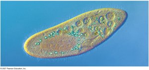
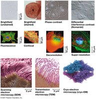
Microscopes are essential tools for visualizing cells and their components, which are typically too small to be seen with the naked eye.
Light Microscopy: Uses visible light and glass lenses to magnify specimens up to 1,000 times.
Electron Microscopy: Includes scanning (SEM) and transmission (TEM) types, providing higher resolution for studying subcellular structures.
Key Parameters:
Magnification: Ratio of image size to actual size.
Resolution: Minimum distance between two distinguishable points.
Contrast: Differences in brightness between parts of the sample.
Cell Fractionation
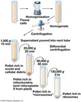
Cell fractionation is a technique used to separate cellular components for study.
Process: Cells are homogenized and then centrifuged at increasing speeds to separate organelles based on size and density.
Purpose: Enables scientists to study the functions of individual organelles.
Prokaryotic vs. Eukaryotic Cells
Basic Features of All Cells
All cells share certain structural features:
Plasma Membrane: Selective barrier for passage of substances.
Cytosol: Semifluid substance within the cell.
Chromosomes: Carry genetic information.
Ribosomes: Sites of protein synthesis.
Prokaryotic Cells
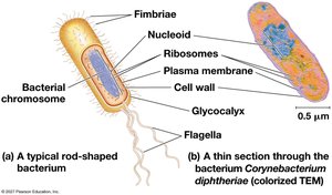
Prokaryotic cells lack a nucleus and membrane-bound organelles.
Nucleoid: Region where DNA is located.
Cell Wall: Provides structural support.
Examples: Bacteria and Archaea.
Eukaryotic Cells
Eukaryotic cells have a nucleus and various membrane-bound organelles.
Nucleus: Contains DNA, surrounded by a double membrane.
Organelles: Specialized structures for different functions.
Size: Generally larger than prokaryotic cells.
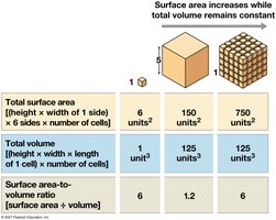
Surface Area to Volume Ratio
The surface area to volume ratio is critical for cell function, affecting the efficiency of material exchange.
As cells increase in size, their volume grows faster than their surface area.
Cells remain small to maintain efficient exchange of materials.
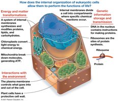
Internal Organization of Eukaryotic Cells
Membranes and Compartmentalization
Eukaryotic cells are divided into compartments by internal membranes, allowing specialized functions to occur simultaneously.
Phospholipid Bilayer: Basic structure of biological membranes.
Organelles: Each compartment provides a unique environment for specific processes.
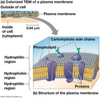
The Plasma Membrane
The plasma membrane is a selectively permeable barrier composed of a phospholipid bilayer with embedded proteins.
Hydrophilic and Hydrophobic Regions: Allow selective passage of substances.
Carbohydrate Side Chains: Play roles in cell recognition and signaling.
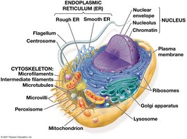
The Nucleus and Ribosomes
The Nucleus
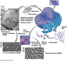
The nucleus is the information center of the cell, housing most of the genetic material.
Nuclear Envelope: Double membrane with nuclear pores for molecular transport.
Chromatin: DNA-protein complex that condenses into chromosomes during cell division.
Nucleolus: Site of ribosomal RNA synthesis.
Ribosomes
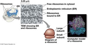
Ribosomes are the sites of protein synthesis, composed of ribosomal RNA and proteins.
Free Ribosomes: Located in the cytosol, synthesize proteins for use within the cell.
Bound Ribosomes: Attached to the endoplasmic reticulum or nuclear envelope, synthesize proteins for secretion or membrane insertion.
The Endomembrane System
Components and Functions
The endomembrane system regulates protein traffic and performs metabolic functions.
Components: Nuclear envelope, endoplasmic reticulum (ER), Golgi apparatus, lysosomes, vacuoles, plasma membrane.
Vesicles: Transport materials between organelles.
Endoplasmic Reticulum (ER)
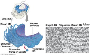
Smooth ER: Synthesizes lipids, detoxifies drugs, stores calcium ions.
Rough ER: Studded with ribosomes, synthesizes proteins, distributes transport vesicles, produces membranes.
Golgi Apparatus
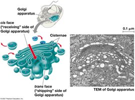
The Golgi apparatus modifies, sorts, and packages proteins and lipids for transport.
Cisternae: Flattened membranous sacs.
cis Face: Receiving side.
trans Face: Shipping side.
Lysosomes
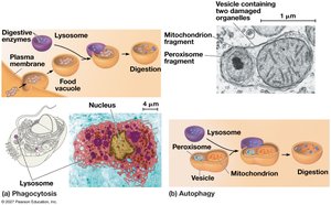
Lysosomes are digestive compartments containing hydrolytic enzymes.
Phagocytosis: Engulfing and digesting foreign particles.
Autophagy: Recycling the cell’s own organelles and macromolecules.
Vacuoles
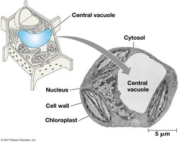
Vacuoles are large vesicles with diverse functions, including storage and maintenance.
Central Vacuole: Found in plant cells, stores inorganic ions and contributes to cell growth.
Contractile Vacuole: Pumps excess water out of cells in protists.
Endomembrane System Review
The endomembrane system is dynamic, coordinating the synthesis, modification, and transport of cellular materials. 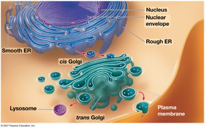
Mitochondria, Chloroplasts, and Peroxisomes
Mitochondria
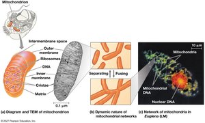
Mitochondria are the sites of cellular respiration, converting energy from nutrients into ATP.
Structure: Double membrane, inner membrane folded into cristae, matrix, intermembrane space.
Function: ATP synthesis, signal integration, synthesis of molecules like melatonin and cortisol.
Chloroplasts
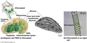
Chloroplasts are the sites of photosynthesis in plants and algae.
Structure: Double membrane, thylakoids stacked into grana, stroma (internal fluid).
Function: Capture light energy and convert it to chemical energy.
Peroxisomes
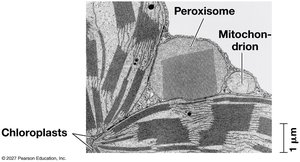
Peroxisomes are oxidative organelles that detoxify harmful substances and break down fatty acids.
Function: Produce hydrogen peroxide, convert fatty acids to sugars, detoxify alcohol.
The Cytoskeleton
Structure and Function
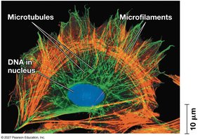
The cytoskeleton is a network of fibers that organizes cell structure and activities.
Components: Microtubules, microfilaments (actin filaments), intermediate filaments.
Functions: Support, motility, transport of organelles, cell division.
Motor Proteins and Cell Motility
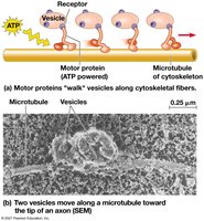
Motor proteins interact with the cytoskeleton to move vesicles and organelles within the cell.
ATP-powered movement: Motor proteins "walk" along cytoskeletal fibers.
Microtubules
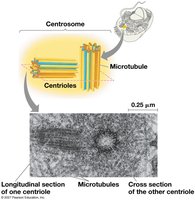
Microtubules are hollow rods made of tubulin, involved in cell shape, organelle movement, and chromosome separation.
Centrosomes and Centrioles: Organize microtubules in animal cells.
Cilia and Flagella
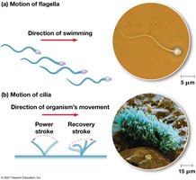
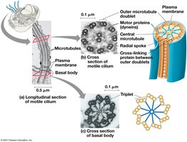
Cilia and flagella are microtubule-containing extensions that enable cell movement.
Structure: Nine doublets of microtubules arranged in a ring with two singles in the center.
Function: Propel cells or move fluid over cell surfaces.
Microfilaments (Actin Filaments)
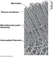
Microfilaments are solid rods of actin, supporting cell shape and enabling movement.
Core of Microvilli: Increase surface area in intestinal cells.
Extracellular Components and Cell Connections
Cell Walls of Plants
Plant cells have cell walls made of cellulose, providing protection and structural support.
Layers: Primary cell wall, middle lamella, secondary cell wall (in some cells).
Extracellular Matrix (ECM) of Animal Cells
Animal cells are surrounded by an extracellular matrix composed of glycoproteins.
Functions: Structural support, cell signaling, regulation of gene activity.
Cell Junctions
Cells in tissues are connected by specialized junctions.
Tight Junctions: Prevent leakage of extracellular fluid.
Desmosomes: Anchor cells together.
Gap Junctions: Allow communication between cells.
Plasmodesmata in Plant Cells
Plasmodesmata are channels that connect plant cells, allowing transport of water, solutes, and signaling molecules.
Summary: Coordination of Cellular Activities
Integration of Cell Components
The cell operates as an integrated unit, with organelles and structures working together to perform essential functions.
Coordination among cytoskeleton, lysosomes, plasma membrane, and other components is vital for processes such as immune response and metabolism.
