 Back
BackA Tour of the Cell: Structure and Function of Eukaryotic and Prokaryotic Cells
Study Guide - Smart Notes
 Tailored notes based on your materials, expanded with key definitions, examples, and context.
A Tour of the Cell
The Fundamental Units of Life
Cells are the basic structural and functional units of all living organisms. They represent the simplest collection of matter that can be alive, and all cells share common features despite their diversity. The study of cells is central to understanding biology, as all organisms are composed of one or more cells.
Cell Theory: All living things are made of cells, and all cells arise from pre-existing cells.
Cell Diversity: Cells can vary greatly in size, shape, and function, but share fundamental characteristics.
Common Features: All cells have a plasma membrane, cytosol, chromosomes, and ribosomes.
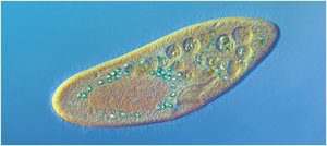
Types of Cells: Prokaryotic vs. Eukaryotic
Cells are classified into two major types: prokaryotic and eukaryotic. This distinction is based on the presence or absence of a nucleus and other membrane-bound organelles.
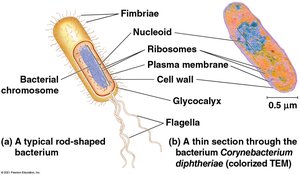
Prokaryotic Cells: Found in Bacteria and Archaea; lack a nucleus and membrane-bound organelles.
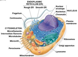
Eukaryotic Cells: Found in Protists, Plants, Animals, and Fungi; possess a nucleus and membrane-bound organelles.
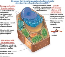
Comparing Prokaryotic and Eukaryotic Cells
Both cell types share certain features, but differ in their internal organization and complexity.
Basic Features of All Cells: Plasma membrane, cytosol, chromosomes, ribosomes.
Prokaryotic Cells:
No nucleus; DNA is located in the nucleoid region.
No membrane-bound organelles.
Smaller size compared to eukaryotic cells.
Eukaryotic Cells:
DNA enclosed within a double-membrane nucleus.
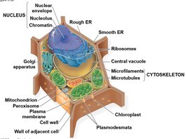
Contains membrane-bound organelles (e.g., mitochondria, ER, Golgi apparatus).
Larger and more complex than prokaryotic cells.
Cell Membranes and Surface Area
Structure of the Plasma Membrane
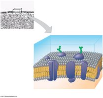
The plasma membrane is a selectively permeable barrier that regulates the movement of substances into and out of the cell. It is composed of a phospholipid bilayer with embedded proteins and carbohydrate side chains.
Phospholipid Bilayer: Hydrophilic heads face outward; hydrophobic tails face inward.
Proteins: Integral and peripheral proteins serve various functions, including transport and signaling.
Carbohydrates: Attached to proteins and lipids, involved in cell recognition.
Surface Area to Volume Ratio
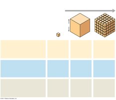
The surface area to volume ratio is a critical factor that limits cell size. As a cell grows, its volume increases faster than its surface area, affecting the efficiency of nutrient and waste exchange.
Formula:
Implication: Smaller cells have a higher ratio, allowing for more efficient exchange with the environment.
The Nucleus and Ribosomes
The Nucleus: Genetic Control Center
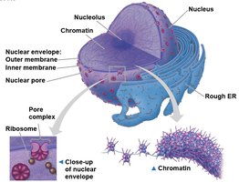
The nucleus is the repository of genetic information in eukaryotic cells. It is surrounded by a double membrane called the nuclear envelope and contains chromatin and the nucleolus.
Nuclear Envelope: Double membrane with nuclear pores for transport.
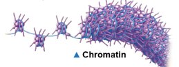
Chromatin: DNA-protein complex; condenses to form chromosomes during cell division.
Nucleolus: Site of ribosomal RNA (rRNA) synthesis.
Ribosomes: Protein Factories
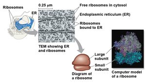
Ribosomes are complexes of rRNA and protein that synthesize polypeptides. They can be free in the cytosol or bound to the endoplasmic reticulum.
Free Ribosomes: Synthesize proteins for use within the cell.
Bound Ribosomes: Synthesize proteins for secretion or for use in membranes.
The Endomembrane System
Components and Functions
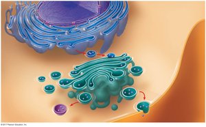
The endomembrane system is a network of membranes within eukaryotic cells that regulates protein traffic and performs metabolic functions.
Components: Nuclear envelope, endoplasmic reticulum (ER), Golgi apparatus, lysosomes, vacuoles, plasma membrane.
Function: Synthesis, modification, sorting, and transport of proteins and lipids.
Endoplasmic Reticulum (ER)
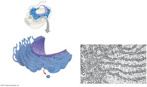
Smooth ER: Synthesizes lipids, metabolizes carbohydrates, detoxifies drugs, stores calcium ions.
Rough ER: Studded with ribosomes; synthesizes proteins, distributes transport vesicles, produces membranes.
Golgi Apparatus
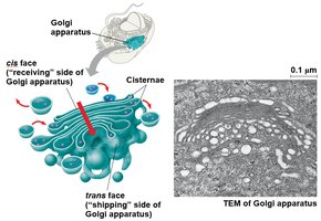
The Golgi apparatus modifies, sorts, and packages products from the ER for secretion or delivery to other organelles.
Cisternae: Flattened membranous sacs.
Cis Face: Receiving side.
Trans Face: Shipping side.
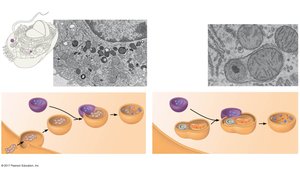
Lysosomes
Lysosomes are membrane-bound sacs containing hydrolytic enzymes for digestion of macromolecules and recycling of cellular components.
Phagocytosis: Digestion of engulfed particles.
Autophagy: Recycling of the cell's own organelles.
Vacuoles
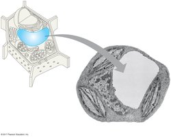
Vacuoles are large vesicles with diverse functions, including storage, waste disposal, and maintaining cell turgor in plants.
Central Vacuole: Prominent in plant cells; stores water and maintains pressure.
Food Vacuole: Formed by phagocytosis.
Contractile Vacuole: Pumps excess water out of cells.
Energy Conversion Organelles
Mitochondria
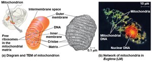
Mitochondria are the sites of cellular respiration, converting chemical energy from food into ATP using oxygen.
Structure: Double membrane, inner membrane folded into cristae, contains its own DNA and ribosomes.
Function: ATP production via oxidative phosphorylation.
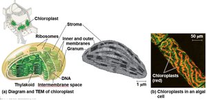
Chloroplasts
Chloroplasts are found in plants and algae and are the sites of photosynthesis, converting solar energy into chemical energy.
Structure: Double membrane, internal thylakoid membranes, contains its own DNA and ribosomes.
Function: Photosynthesis:
The Cytoskeleton
Structure and Function
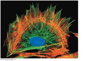
The cytoskeleton is a dynamic network of protein fibers that provides structural support, organizes cell contents, and enables cell movement.
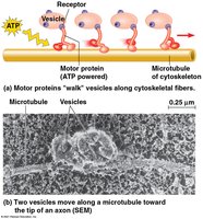
Microtubules: Hollow tubes; maintain cell shape, guide organelle movement, separate chromosomes.
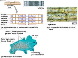
Microfilaments (Actin Filaments): Solid rods; support cell shape, involved in muscle contraction and cell motility.
Intermediate Filaments: Fibrous proteins; maintain cell shape, anchor organelles.
Roles in Cell Motility
Motor Proteins: Move vesicles and organelles along cytoskeletal tracks using ATP.
Cell Movement: Includes muscle contraction, cytoplasmic streaming, and amoeboid movement.
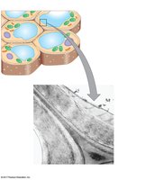
Extracellular Components and Cell Connections
Cell Walls of Plants
Plant cells are surrounded by a rigid cell wall composed of cellulose, which provides structural support and protection.
Primary Cell Wall: Thin and flexible.
Secondary Cell Wall: Thicker and stronger, formed in some cells.
Plasmodesmata: Channels that connect plant cells for communication.
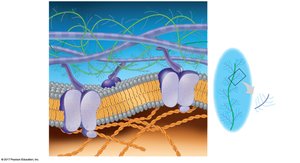
Extracellular Matrix (ECM) of Animal Cells
The ECM is a complex network of glycoproteins (collagen, proteoglycans, fibronectin) that provides structural support and mediates cell signaling in animal tissues.
Integrins: Membrane proteins that connect the ECM to the cytoskeleton.
Function: Regulates cell behavior, adhesion, and communication.
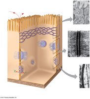
Cell Junctions in Animal Cells
Animal cells are connected by specialized junctions that facilitate communication and maintain tissue integrity.
Tight Junctions: Prevent leakage of extracellular fluid.
Desmosomes: Anchor cells together via intermediate filaments.
Gap Junctions: Allow passage of ions and small molecules between cells.
Junction Type | Structure | Function |
|---|---|---|
Tight Junction | Membranes pressed together | Seals cells, prevents leakage |
Desmosome | Intermediate filaments | Anchors cells together |
Gap Junction | Channels between cells | Communication |
Summary
This chapter provides a comprehensive overview of cell structure and function, highlighting the differences between prokaryotic and eukaryotic cells, the organization of cellular components, and the roles of membranes, organelles, and the cytoskeleton in maintaining life processes.
