 Back
BackAnatomy of the Thorax and Abdomen: The Skeleton, Joints, and Ligaments
Study Guide - Smart Notes
 Tailored notes based on your materials, expanded with key definitions, examples, and context.
The Skeleton of the Thorax and Abdomen
The Vertebral Column
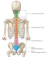
The vertebral column forms the central axis of the skeleton and is essential for structural support, movement, and protection of the spinal cord. It consists of vertebrae and intervertebral disks, organized into distinct regions.
Vertebrae: There are approximately 33 vertebrae, divided into 7 cervical, 12 thoracic, 5 lumbar, 5 sacral (fused), and 3–4 coccygeal (fused).
Mnemonic: "You eat breakfast at 7, lunch at 12, and dinner at 5" helps recall the number of vertebrae in each region.
Curvatures: The column has three major curvatures: cervical lordosis, thoracic kyphosis, and lumbar lordosis.
Clinical Correlation: Excessive thoracic curvature leads to kyphosis (hunchback), excessive lumbar curvature to lordosis (swayback), and excessive lateral curvature to scoliosis.
Structure of a Typical Vertebra
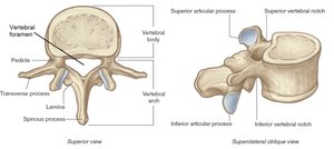
A typical vertebra is composed of several key anatomical features that facilitate its function and articulation.
Vertebral body: Weight-bearing portion, linked to adjacent vertebrae by intervertebral disks and ligaments.
Vertebral foramen: Forms the vertebral canal, which houses the spinal cord.
Vertebral arch: Lateral and posterior part of the vertebral foramen.
Pedicle: Bony pillars attaching the arch to the body.
Laminae: Flat sheets forming the roof of the arch.
Spinous and transverse processes: Sites for muscle and ligament attachment; transverse processes in thoracic vertebrae are sites for rib attachment.
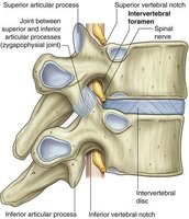
Articular processes: Superior and inferior, for articulation with adjacent vertebrae.
Vertebral notches: Superior and inferior, participate in formation of intervertebral foramina for spinal nerves.
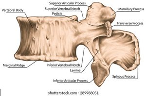
Vertebral Segments
Cervical Vertebrae
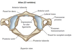
The cervical region contains seven vertebrae, characterized by transverse foramina. The first two, the Atlas (C1) and Axis (C2), are specialized for head movement.
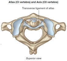
Atlas (C1): Lacks a vertebral body; permits nodding of the head at the atlanto-occipital joint. The dens of C2 is held by the transverse ligament of the atlas.
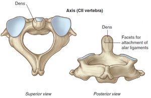
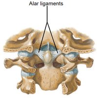
Axis (C2): Contains the dens, which acts as a pivot for head rotation. Alar ligaments limit excessive rotation.
C7: Vertebra prominens, easily palpable.
Thoracic Vertebrae
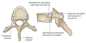
There are twelve thoracic vertebrae, all of which articulate with ribs. They have demifacets and facets for rib articulation.
Facets: On bodies for heads of ribs; on transverse processes for tubercles of ribs.
Lumbar Vertebrae
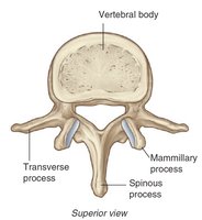
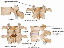
The five lumbar vertebrae are large and lack facets for rib articulation. L5 has the largest vertebral body, supporting the most weight. Interlaminar spaces allow for lumbar puncture procedures.
Sacral Vertebrae
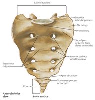
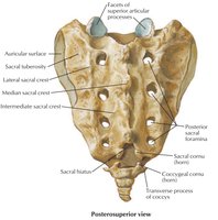
The sacrum is a single bone formed by the fusion of five vertebrae. It articulates with the os coxae via auricular processes and contains sacral foramina for passage of spinal nerves. The sacral promontory is a key landmark in obstetrics.
Coccygeal Vertebrae
The coccyx is a small triangular bone formed by 3–4 fused vertebrae, representing the remnant of caudal vertebrae in vertebrates.
The Sternum
Anatomy of the Sternum
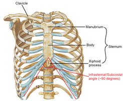
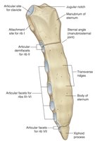
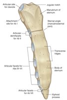
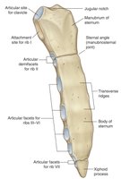
The sternum is a flat bone in the center of the thorax, consisting of three parts: the manubrium, the body, and the xiphoid process. It serves as an attachment point for ribs and clavicles.
Manubrium: Forms part of the neck and thorax framework; has jugular notch, articulates with clavicles and 1st costal cartilages, and contains a demifacet for the 2nd costal cartilage. The sternal angle is a key landmark.
Body: Flat, marked by transverse ridges; lateral margins have articular facets for costal cartilages; attached to the xiphoid process.
Xiphoid process: Smallest, variable shape; begins cartilaginous and ossifies in adulthood; articulates with the 7th costal cartilage.
The Ribs
Classification and Structure
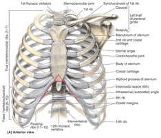
The ribs are twelve pairs of bones terminating anteriorly as costal cartilages. They are classified as true, false, and floating ribs based on their anterior connections.
True ribs: Upper seven pairs; costal cartilages articulate directly with the sternum.
False ribs: Lower five pairs; costal cartilages of VIII–X articulate with those above; XI and XII are floating ribs with no anterior connection.
Typical Rib Anatomy
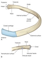
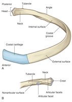
A typical rib is curved, with an anterior end continuous with costal cartilage and a posterior end articulating with the vertebral column. Key landmarks include the head, neck, tubercle, shaft, and costal groove.
Head: Has two articular surfaces for articulation with vertebral bodies.
Neck: Separates head from tubercle.
Tubercle: Articular portion for transverse process; non-articular for ligament attachment.
Shaft: Thin, flat, bends at the angle; costal groove accommodates intercostal vessels and nerve.
Clinical correlation: In pleurocentesis, the needle is inserted just above the upper edge of a rib to avoid the neurovascular bundle in the costal groove.
Atypical Ribs
Some ribs have unique features and are classified as atypical: I, II, X, XI, and XII.
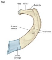
Rib I: Flat, broad surfaces; head has one articular surface; distinct scalene tubercle separates grooves for subclavian vessels.
Rib II: Flat, twice as long as Rib I.
Rib X: Single facet for articulation with T10.
Ribs XI and XII: Articulate only with vertebral bodies; no tubercles or necks; short and pointed anteriorly.
Joints of the Trunk
Joints Between Vertebrae
Vertebral joints include symphyses and synovial joints, providing stability and flexibility.
Intervertebral disks: Symphysis between vertebral bodies; composed of annulus fibrosus (outer ring) and nucleus pulposus (gelatinous center).
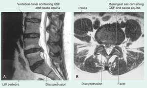
Clinical correlation: Herniation of nucleus pulposus can impinge on spinal nerve roots, causing a herniated disk.
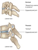
Zygapophysial joints: Synovial joints between articular processes.
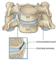
Uncovertebral joints: Only in cervical spine; between uncinate process and body of superior vertebra.
Joints Between Ribs and Vertebral Column
These joints allow for rib movement during respiration.
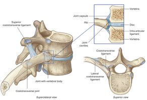
Costovertebral joints: Between rib heads and vertebral bodies.
Costotransverse joints: Between rib tubercles and transverse processes.
Function: Allow rotation and elevation/depression of ribs, altering thoracic cavity dimensions.
Joints of Anterior Ribs and Sternum
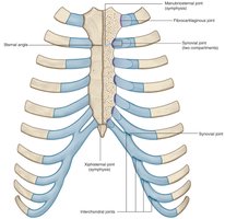
Sternocostal joints: Between upper 7 costal cartilages and sternum.
Interchondral joints: Between costal cartilages of ribs VII–X.
Manubriosternal joint: Between manubrium and body; sternal angle is a key landmark for rib counting and clinical procedures.
Xiphisternal joint: Between body and xiphoid process.
Ligaments of the Vertebral Column
Major Ligaments
Ligaments stabilize the vertebral column and limit excessive movement.
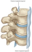
Anterior and posterior longitudinal ligaments: Run along the length of the vertebral column, limiting extension and flexion.
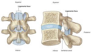
Ligamenta flava: Connect adjacent laminae, preserving upright posture.
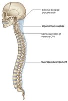
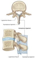
Supraspinous ligament and ligamentum nuchae: Connect spinous processes; ligamentum nuchae is prominent in the cervical region.
Interspinous ligaments: Connect adjacent spinous processes.
Summary Table: Vertebral Column Regions
Region | Number of Vertebrae | Key Features |
|---|---|---|
Cervical | 7 | Transverse foramina, Atlas and Axis specialized |
Thoracic | 12 | Articulate with ribs, demifacets |
Lumbar | 5 | Large bodies, no rib facets |
Sacral | 5 (fused) | Articulates with pelvis, sacral foramina |
Coccygeal | 3–4 (fused) | Small, vestigial |
Summary Table: Rib Classification
Type | Number | Anterior Connection |
|---|---|---|
True | 1–7 | Directly to sternum |
False | 8–10 | Indirect, via costal cartilage |
Floating | 11–12 | None |
