 Back
BackAnimal Development: Fertilization, Cleavage, Morphogenesis, and Cell Fate Specification
Study Guide - Smart Notes
 Tailored notes based on your materials, expanded with key definitions, examples, and context.
CHAPTER 47: ANIMAL DEVELOPMENT
Overview of Animal Development
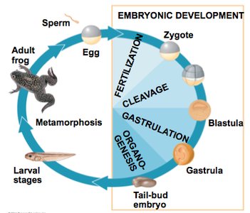
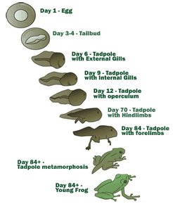
Animal development is a complex, multi-stage process that transforms a single fertilized cell into a fully formed organism. The main stages include fertilization, cleavage, gastrulation, organogenesis, and metamorphosis. Model organisms such as sea urchins, frogs, chicks, and nematodes are commonly used to study these processes due to their accessibility and experimental tractability.
Fertilization and Cleavage
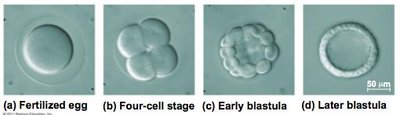
Fertilization is the process by which haploid gametes (sperm and egg) fuse to form a diploid zygote. This event initiates embryonic development and is followed by cleavage, a series of rapid cell divisions without growth, partitioning the cytoplasm into smaller cells called blastomeres.
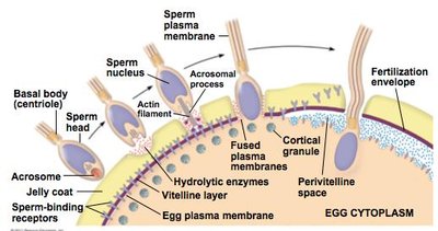
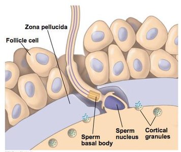
Fertilization Sequence: Sperm penetrates the egg's protective layer, egg surface receptors bind sperm molecules, and changes at the egg surface prevent polyspermy.
Polyspermy Block: Fast block (membrane depolarization) and slow block (cortical reaction, Ca2+ elevation) prevent multiple sperm from fertilizing the egg.
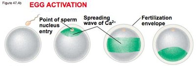
Egg Activation: Ca2+ influx increases cellular respiration and protein synthesis, activating the egg and initiating cell division.
Cleavage and Blastula Formation
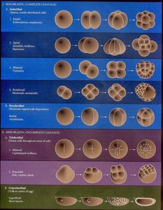
Cleavage divides the zygote into blastomeres, forming a blastula with a fluid-filled cavity called the blastocoel. The pattern of cleavage is influenced by yolk distribution.
Holoblastic Cleavage: Complete division in eggs with little yolk (e.g., mammals, frogs).
Meroblastic Cleavage: Incomplete division in yolk-rich eggs (e.g., reptiles, fish).
Morphogenesis: Gastrulation and Organogenesis
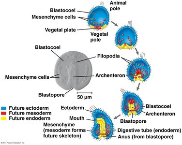
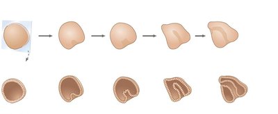
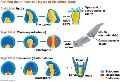
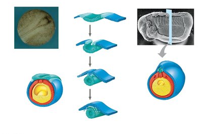
Morphogenesis is the process by which cells migrate and rearrange to form the three primary germ layers during gastrulation, followed by organogenesis, the formation of organs.
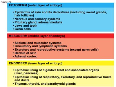
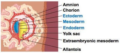
Gastrulation: Rearranges blastula cells into a gastrula with ectoderm, mesoderm, and endoderm.
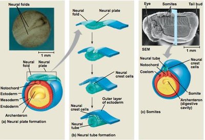
Organogenesis: Germ layers develop into rudimentary organs; notochord forms from mesoderm, neural plate from ectoderm.
Gastrulation in Humans
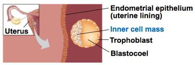
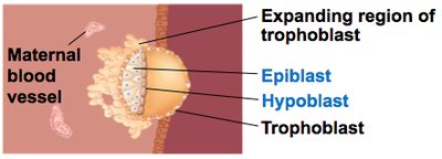
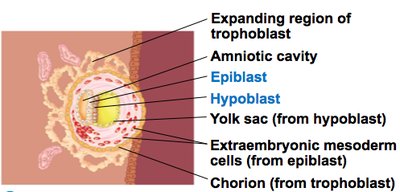
Human eggs have little yolk, and the blastocyst is the equivalent of the blastula. The trophoblast initiates implantation, and extra-embryonic membranes form as gastrulation begins.
Blastocyst: Inner cell mass forms the embryo; trophoblast initiates implantation.
Extra-embryonic Membranes: Chorion (gas exchange), amnion (fluid enclosure), yolk sac (blood cell formation), allantois (waste disposal).
Organogenesis: Formation of Notochord and Neural Tube
Organogenesis involves the development of organs from the three germ layers. In vertebrates, the notochord forms from mesoderm, and the neural plate forms from ectoderm, eventually giving rise to the neural tube and central nervous system.
Neural Crest Cells: Migrate to form nerves, teeth, and skull bones.
Somites: Mesodermal blocks that form vertebrae, ribs, and associated muscles.
The Cytoskeleton and Programmed Cell Death
The cytoskeleton, composed of microtubules and microfilaments, is essential for cell shape changes and migration during development. Programmed cell death (apoptosis) eliminates excess cells, such as neurons, to refine tissue structure.
Cytoplasmic Determinants and Inductive Signals
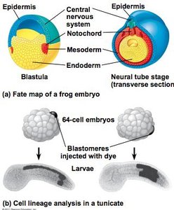
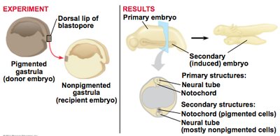
Cell fate specification is governed by cytoplasmic determinants and inductive signals. Determination commits cells to a specific fate, while differentiation results in specialized structure and function. Inductive signals from neighboring cells influence pattern formation and spatial organization.
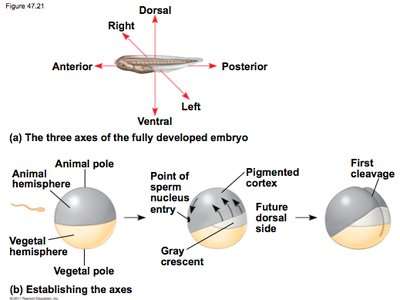
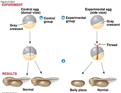
Axis Formation: Anterior-posterior and dorsal-ventral axes are established by cytoplasmic determinants and fertilization events.
Totipotency: Early blastomeres can develop into all cell types.
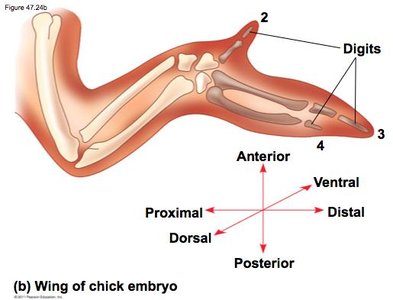
Pattern Formation: Positional information guides cells in limb development along proximal-distal, anterior-posterior, and dorsal-ventral axes.
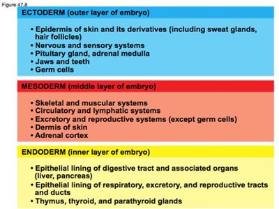
Summary Table: Germ Layer Derivatives
Germ Layer | Derivatives |
|---|---|
Ectoderm | Epidermis, nervous system, pituitary gland, jaws, teeth, germ cells |
Mesoderm | Skeletal, muscular, circulatory, lymphatic, excretory, reproductive systems, dermis, adrenal cortex |
Endoderm | Digestive tract lining, liver, pancreas, respiratory, excretory, reproductive tract lining, thymus, thyroid, parathyroid glands |
Example: The notochord forms from mesoderm and acts as a signaling center for neural tube development, which arises from ectoderm.
Additional info: The notes have been expanded to include definitions, examples, and context for each developmental stage, as well as a summary table of germ layer derivatives.
