 Back
BackAnimal Diversity and Dissection Study Guide: Opisthokonta, Fungi, Lophotrochozoa, Ecdysozoa, and Deuterostomia
Study Guide - Smart Notes
 Tailored notes based on your materials, expanded with key definitions, examples, and context.
Opisthokonta
Overview of Opisthokonta
The Opisthokonta is a broad eukaryotic clade that includes both the animal and fungal kingdoms, as well as several related protist groups. This group is characterized by the presence of a single posterior flagellum in the motile cells of many of its members.
Key Members: Animals (Metazoa), Fungi, and choanoflagellates.
Choanoflagellates: Unicellular or colonial protists considered the closest living relatives of animals. Their collar cells resemble the choanocytes of sponges, supporting the evolutionary link between these groups.
Significance: Understanding Opisthokonta helps clarify the evolutionary origins of multicellularity and animal development.
Fungi, Parazoa, & Radiata
Fungi: Structure and Symbiosis
Fungi are eukaryotic organisms that play essential roles in ecosystems as decomposers, symbionts, and pathogens. The Basidiomycota is a major fungal phylum known for producing large fruiting bodies (basidiocarps).
Generalized Anatomy of Basidiomycota: Includes cap, gills, stalk, and mycelium.
Symbiosis: Many fungi form mutualistic relationships with plants (mycorrhizae), enhancing nutrient uptake.
Lichens: Symbiotic associations between fungi (usually Ascomycota) and photosynthetic partners (algae or cyanobacteria). Lichens exhibit various growth forms: crustose, foliose, and fruticose.
Parazoa and Radiata
Parazoa includes sponges (Phylum Porifera), which lack true tissues and organs. Radiata includes cnidarians and ctenophores, which exhibit radial symmetry and have true tissues but lack bilateral symmetry.
Sponges: Characterized by a porous body and choanocyte-lined chambers for filter feeding.
Radiata: Includes jellyfish, corals, and sea anemones, with a gastrovascular cavity and specialized stinging cells (cnidocytes).
Metazoa
Generalized Anatomy and Classification of Sponges
Sponges are simple multicellular animals with specialized cells but no true tissues. They are classified based on their skeletal structure (spicules) and canal systems.
Key Structures: Osculum, spongocoel, choanocytes, and spicules.
Classification: Based on body plan (asconoid, syconoid, leuconoid) and spicule composition (calcareous, siliceous, or spongin).
Lophotrochozoa
Anatomical Structures and Representative Organisms
Lophotrochozoa is a diverse clade of protostome animals, including flatworms, mollusks, and annelids. Classification is based on the presence of a lophophore (feeding structure) or trochophore larva.
Tapeworm Anatomy: Scolex (head), proglottids (body segments), and hooks/suckers for attachment.
Earthworm Anatomy: Segmented body, clitellum, setae, and a complete digestive tract.
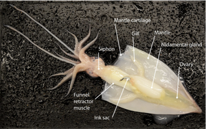
Squid Anatomy: Mantle, siphon, arms, tentacles, beak, ink sac, and internal shell (pen).
Classification: Based on body plan, segmentation, and larval forms.
Ecdysozoa
Arthropod Anatomy and Classification
Ecdysozoa includes animals that molt their exoskeleton, such as arthropods and nematodes. Key arthropod groups include insects, crustaceans, and myriapods.
Horseshoe Crab, Crayfish, and Grasshopper Anatomy: Segmented body, jointed appendages, exoskeleton, and specialized mouthparts.
Distinguishing Insecta: Three body segments (head, thorax, abdomen), three pairs of legs, and usually two pairs of wings.
Chilopoda vs. Diplopoda: Centipedes (Chilopoda) have one pair of legs per segment; millipedes (Diplopoda) have two pairs per segment.
Deuterostomia
Chordate and Echinoderm Characteristics
Deuterostomia includes echinoderms and chordates, united by embryonic development patterns (blastopore becomes anus).
Chordate Characteristics: Notochord, dorsal hollow nerve cord, pharyngeal slits, and post-anal tail.
Mammal Characteristics: Hair, mammary glands, three middle ear bones, and a neocortex.
Sea Star Anatomy: Central disc, arms (rays), madreporite, tube feet, and water vascular system.
Sea Star Development: Radial cleavage, blastula, gastrula, and larval stages.
Mammalian Dentition: Differentiated teeth (incisors, canines, premolars, molars) adapted for varied diets.
Representative Dissection Images
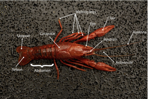
Crayfish Dissection
The crayfish is a representative crustacean used to study arthropod anatomy. Key external features include the carapace, abdomen, walking legs, claws, antennae, and specialized tail structures (telson and uropods).
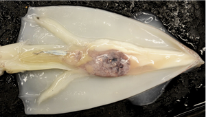
Squid Dissection
Squid are cephalopod mollusks with specialized adaptations for predation and locomotion. Dissection reveals the internal arrangement of organs, including the mantle, gills, ink sac, and reproductive structures.
Summary Table: Major Animal Groups and Key Features
Group | Key Characteristics | Representative Example |
|---|---|---|
Opisthokonta | Posterior flagellum, includes animals and fungi | Choanoflagellate, Homo sapiens |
Fungi | Chitin cell walls, decomposers, symbionts | Mushroom (Basidiomycota) |
Lophotrochozoa | Lophophore/trochophore larva, diverse body plans | Earthworm, Squid |
Ecdysozoa | Molting (ecdysis), exoskeleton | Crayfish, Grasshopper |
Deuterostomia | Blastopore forms anus, radial cleavage | Sea star, Mammal |
Additional info: These notes synthesize key anatomical and phylogenetic concepts relevant to the major animal groups and their representative laboratory specimens, as outlined in the provided study guide. For detailed dissections and further examples, refer to your lab manual and textbook chapters on animal diversity and anatomy.
