 Back
BackTopic 3 - Animal Form & Function
Study Guide - Smart Notes
 Tailored notes based on your materials, expanded with key definitions, examples, and context.
Animal Form and Function
Introduction to Animal Tissues
Animals are composed of specialized cells organized into tissues, which perform distinct functions necessary for survival. The adult animal body is derived from three embryonic germ layers and consists of four primary tissue types: epithelial, muscle, nervous, and connective tissues. Each tissue type has unique structural and functional characteristics that contribute to the overall physiology of the organism.
Adult Tissues
Epithelial Tissue
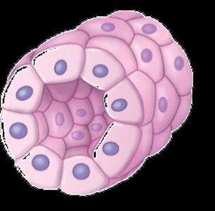
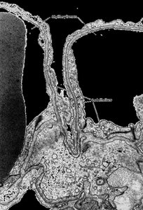
Epithelial tissue forms the outer coverings of the body and lines internal tubes and cavities. It consists of one or more layers of tightly packed cells resting on a basement membrane. Epithelia serve as barriers, control the movement of substances, and are involved in absorption and secretion.
Protection: Acts as a physical barrier against mechanical injury, pathogens, and fluid loss.
Control of Movement: Regulates diffusion, facilitated diffusion, and active transport of substances.
Absorption & Secretion: Specialized for uptake of nutrients and release of products such as mucus or enzymes.
Specialized Connections in Epithelia
Gap Junctions: Allow communication between adjacent cells.
Tight Junctions: Prevent leakage of molecules between cells and restrict migration of membrane proteins.
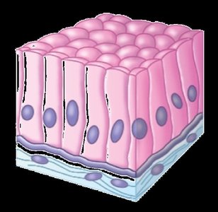
Polarity: Epithelial cells have an apical surface (facing the lumen or outside) and a basolateral surface (facing underlying tissues).
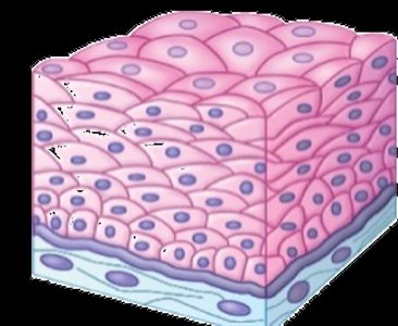
Classification of Epithelia
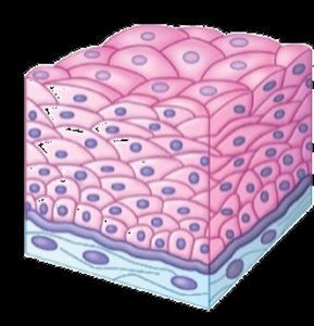
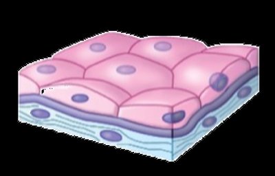
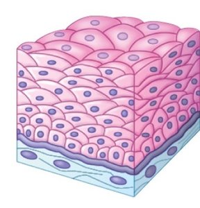
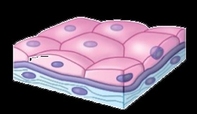
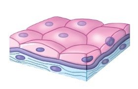
By Layers: Simple (one layer) or stratified (multiple layers).
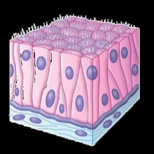
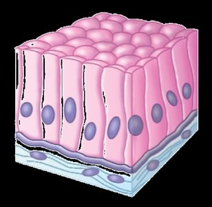
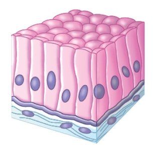
By Cell Shape: Squamous (flat), cuboidal (cube-shaped), columnar (tall and column-like).
Type | Layers | Shape | Main Function |
|---|---|---|---|
Simple Squamous | 1 | Flat | Diffusion/filtration |
Simple Cuboidal | 1 | Cube | Secretion/absorption |
Simple Columnar | 1 | Column | Absorption/secretion |
Stratified Squamous | Multiple | Flat | Protection |

Structure versus Function in Epithelia
Gas Exchange: Simple squamous epithelia in lungs and gills facilitate efficient diffusion of gases.
Absorption: Columnar epithelia in the small intestine maximize nutrient uptake and provide some protection.
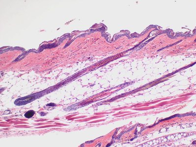
Protection: Stratified squamous epithelia in the epidermis protect against abrasion and dehydration.

Muscle Tissue
Muscle tissue is specialized for contraction and movement. It is excitable and contractile, allowing animals to move and manipulate their environment. There are three main types:
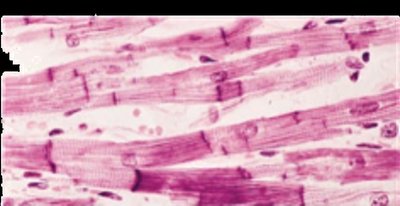
Skeletal Muscle: Voluntary, striated muscle attached to bones for movement.
Cardiac Muscle: Involuntary, striated muscle found only in the heart.
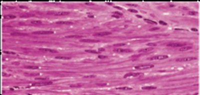
Smooth Muscle: Involuntary, non-striated muscle found in walls of internal organs.
Structure versus Function in Muscle Tissue
Skeletal and Cardiac Muscle: Both are striated due to the regular arrangement of actin and myosin proteins into sarcomeres. This organization allows for powerful, coordinated contractions.
Sarcomeres: The basic contractile unit; shortening of many sarcomeres in series results in overall muscle contraction.
Smooth Muscle: Lacks striations; actin and myosin are present but less organized. Contractions are slower and can be involuntary (in vertebrates) or voluntary (in invertebrates).
Nervous Tissue
Nervous tissue is specialized for the conduction of electrical impulses. It consists of two main cell types:
Neurons: Excitable cells that transmit nerve impulses.
Glia: Supportive cells that nourish, insulate, and protect neurons.
Connective Tissue
Connective tissue supports, binds, and protects other tissues and organs. It is characterized by cells scattered within an abundant extracellular matrix, which may be liquid, gel-like, or solid.
Examples: Bone (rigid support), cartilage, adipose (fat), blood, tendons, and ligaments.
Function: Provides structural support, stores energy, transports substances, and protects organs.
Tissues, Organs, and Organ Systems
Tissue Organization in Organs
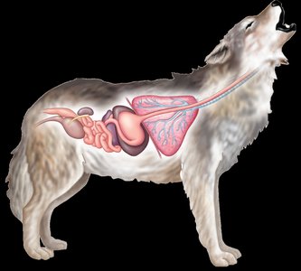
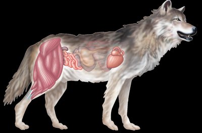
Organs are structures composed of two or more tissue types working together to perform specific functions. For example, the small intestine contains all four adult tissue types: epithelial (lining), muscle (movement), nervous (control), and connective (support).
Organ Systems
Organ systems are groups of organs that work together to carry out major body functions. Division of labor among systems increases efficiency and specialization.
Internal versus External Environments
Animals maintain internal environments that may differ significantly from their external surroundings. Key parameters regulated include concentration of solutes, pH, and temperature. Exchange of compounds, ions, and heat occurs across body surfaces.
Regulation or Conformation
Animals may either regulate internal parameters (regulators) or allow them to conform to the environment (conformers). For example, some fish maintain constant internal osmolarity, while others match their internal environment to seawater.
Homeostasis
Homeostasis is the active maintenance of a stable internal environment despite external fluctuations. It involves physiological processes that keep parameters such as temperature, pH, and osmolarity within narrow limits.
Homeothermy vs. Poikilothermy: Homeotherms maintain a constant body temperature; poikilotherms allow body temperature to vary with the environment.
Endothermy vs. Ectothermy: Endotherms generate heat metabolically; ectotherms rely on external sources for heat.
Mechanisms of Homeostasis
Thermoregulation: The hypothalamus acts as a thermostat, sensing body temperature, comparing it to a setpoint, and initiating corrective responses.
Adaptations to Conserve Heat: Insulation (hair, feathers, fat) and vascular heat exchangers help retain body heat.
Osmoregulation and Osmoconformation
Osmoregulation is the control of water and solute concentrations in the body. Animals must balance water gain and loss, especially in aquatic environments with varying osmolarities (e.g., seawater vs. freshwater).
Sites of Water Exchange: Gills, gut, skin, and kidneys are major sites for water and ion movement.
Osmolarity: Seawater (~1000 mOsm), freshwater (5–50 mOsm), and vertebrate body fluids (~330 mOsm) require different osmoregulatory strategies.
Example: Marine fish lose water to the environment and must drink seawater, excreting excess salts; freshwater fish gain water and must excrete dilute urine.
Self-Guided Learning Tips
Understand the structure and function of each tissue type.
Be able to visually or descriptively identify tissue subtypes.
Relate tissue structure to its function in the body.
Classify tissues (e.g., bone as a connective tissue) and distinguish them from others based on unique features.
