 Back
Animal Movement: Muscle and Skeletal Systems
Study Guide - Smart Notes
 Tailored notes based on your materials, expanded with key definitions, examples, and context.
Animal Movement
Introduction to Animal Movement
Animal movement is a fundamental biological process that enables organisms to respond to environmental stimuli. Movement can involve the entire organism (locomotion) or specific body parts relative to the whole. The muscle and skeletal systems work together to produce coordinated movement, which is essential for survival, foraging, escaping predators, and reproduction.
Muscle Structure and Function
Muscle Organization
Muscle tissue is organized into bundles of muscle fibers, each fiber being a single cell containing many myofibrils. Myofibrils are composed of repeating units called sarcomeres, which are the fundamental contractile units of muscle.
Muscle fiber: A single, elongated cell containing multiple myofibrils.
Myofibril: Composed of repeating sarcomeres, giving muscle its striated appearance.
Sarcomere: The basic contractile unit, defined by the region between two Z-disks.
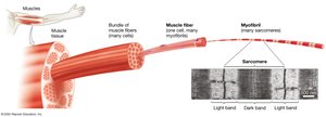
Sarcomere Structure
Sarcomeres are made up of two main types of protein filaments:
Thin filaments: Primarily composed of actin, anchored to the Z-disk.
Thick filaments: Composed of myosin molecules.
The arrangement of these filaments creates the characteristic light and dark bands seen in striated muscle.
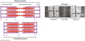
Sliding Filament Model of Muscle Contraction
Muscle contraction occurs through the sliding of thin (actin) filaments past thick (myosin) filaments, shortening the sarcomere without changing the length of the filaments themselves. This process is powered by ATP and is known as the sliding filament model.
Myosin heads: Bind to actin and use ATP hydrolysis to generate force.
Actin-myosin interaction: Repeated cycles of attachment, pivoting, and detachment pull the Z-disks closer together.
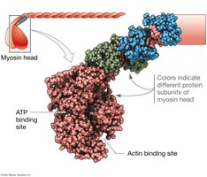
Steps of the Sliding Filament Cycle
ATP binds to myosin head, causing it to release actin.
ATP hydrolysis "cocks" the myosin head into a high-energy state.
Myosin binds to a new position on actin, forming a cross-bridge.
Release of inorganic phosphate (Pi) initiates the power stroke, moving actin.
ADP is released, and the cycle is ready to repeat.
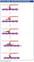
Regulation of Muscle Contraction
Role of Regulatory Proteins
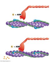
Muscle contraction is regulated by the proteins tropomyosin and troponin on the thin filament. In a relaxed muscle, these proteins block the myosin-binding sites on actin, preventing contraction.
Tropomyosin: Covers myosin-binding sites on actin.
Troponin: Binds calcium ions (Ca2+), causing a conformational change that moves tropomyosin away from binding sites.
Calcium and Muscle Activation
When a muscle is stimulated by a nerve impulse, Ca2+ is released from the sarcoplasmic reticulum. Ca2+ binds to troponin, shifting the troponin-tropomyosin complex and exposing myosin-binding sites on actin, allowing contraction to proceed.
Neural Control of Muscle Contraction
Neuromuscular Junction and Excitation-Contraction Coupling
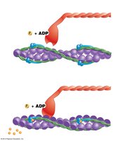
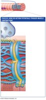
Muscle contraction is initiated by action potentials from motor neurons. The sequence is as follows:
An action potential arrives at the motor neuron's axon terminal, releasing acetylcholine (ACh).
ACh binds to receptors on the muscle cell membrane, generating an action potential in the muscle fiber.
The action potential travels along the membrane and into T-tubules, triggering Ca2+ release from the sarcoplasmic reticulum.
Ca2+ binds to troponin, initiating contraction.
Types of Muscle Tissue
Classification and Characteristics
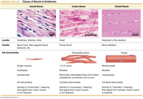
There are three main types of muscle tissue in animals:
Skeletal muscle: Voluntary, striated, attached to bones, responsible for body movement.
Cardiac muscle: Involuntary, striated, found in the heart, responsible for pumping blood.
Smooth muscle: Involuntary, non-striated, found in internal organs, responsible for movements like peristalsis.
Type | Location | Function | Cell Characteristics |
|---|---|---|---|
Smooth | Intestines, arteries | Move food, regulate blood pressure | Single nucleus, unstriated, involuntary |
Cardiac | Heart | Pump blood | 1-2 nuclei, striated, involuntary, intercalated discs |
Skeletal | Attached to skeleton | Move skeleton | Multinucleate, striated, voluntary |
Skeletal Muscle Fiber Types and Organization
Fiber Types
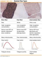
Skeletal muscle fibers are classified based on their contraction speed, metabolic pathway, and fatigue resistance:
Type | Color | ATP Source | Mitochondria | Fatigue |
|---|---|---|---|---|
Slow | Red | Oxidative phosphorylation | Many | Fatigue slowly |
Fast | White | Glycolysis | Few | Fatigue quickly |
Intermediate | Pink or red | Glycolysis and oxidative phosphorylation | Many | Intermediate fatigue |
Muscle Fiber Arrangement
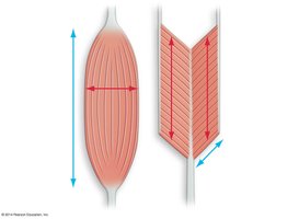
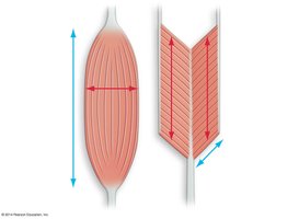
The arrangement of muscle fibers affects force and movement:
Parallel fibers: Maximize length change (e.g., sartorius muscle).
Pennate fibers: Maximize force due to greater cross-sectional area (e.g., gastrocnemius muscle).
Skeletal Systems
Types of Skeletal Systems
Skeletal systems provide support, protection, and leverage for movement. There are three main types:
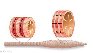
Hydrostatic skeletons: Use fluid pressure (e.g., earthworms).
Endoskeletons: Internal hard structures (e.g., vertebrates).
Exoskeletons: External hard coverings (e.g., arthropods).
Endoskeleton Structure and Function
Vertebrate endoskeletons are composed of:
Cartilage: Cushions joints.
Bone: Provides rigid support and movement (206 bones in adult humans).
Ligaments: Connect bones to bones.
Tendons: Connect muscles to bones.
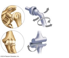
Muscle and Joint Interaction
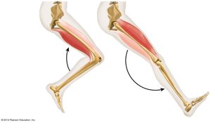
Movement is produced by antagonistic muscle pairs (flexors and extensors) acting across joints. Muscles can only pull, not push, so coordinated contraction and relaxation are necessary for movement.
Exoskeletons
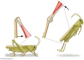
Exoskeletons are rigid external coverings that provide protection and points of muscle attachment. Insects have exoskeletons made of chitin, while crustaceans have mineralized cuticles. Movement is achieved by paired flexor and extensor muscles acting on joints.
Locomotion
Modes and Forces of Locomotion
Animals move in various ways depending on their environment:
Land: Gravitational and inertial forces dominate.
Water: Drag forces are significant.
Air: Body weight, gravity, and drag must be overcome.
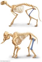
Body size, shape, and posture influence locomotion efficiency. Natural selection favors minimizing the energetic cost of movement.
Cost of Locomotion
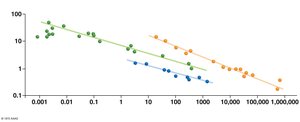
The energy cost of locomotion varies by mode:
Running is most costly per unit distance, followed by flying, then swimming.
Larger animals are generally more efficient movers than smaller animals.
Additional info: This guide covers the structure and function of muscle and skeletal systems, the molecular basis of muscle contraction, and the principles of animal locomotion, integrating key concepts from animal physiology and biomechanics.
