 Back
Animal Nutrition: Structure, Function, and Homeostasis
Study Guide - Smart Notes
 Tailored notes based on your materials, expanded with key definitions, examples, and context.
Animal Nutrition
Introduction to Animal Nutrition
Animal nutrition is a fundamental topic in biology, focusing on how animals obtain and process nutrients to sustain life. Animals require chemical energy for ATP synthesis and carbon-containing compounds for building molecules. As heterotrophs, animals must acquire these nutrients from other organisms.
Four key processes:
Ingestion: Intake of food into the digestive tract.
Digestion: Breakdown of food into smaller molecules.
Absorption: Uptake of nutrients across the digestive epithelium.
Elimination: Removal of waste from the digestive tract.
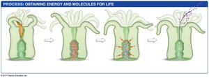
Nutritional Requirements
Animals obtain energy and building blocks from carbohydrates, proteins, and fats. These are classified as nutrients, substances essential for life. The energy content of food is measured in kilocalories (Calories).
Essential nutrients: Cannot be synthesized by the organism and must be obtained from the diet. Includes essential amino acids, essential fatty acids, vitamins, and minerals.
Vitamins: Organic compounds required in small amounts, often functioning as coenzymes.
Minerals: Inorganic substances used as enzyme cofactors, structural materials, and electrolytes.
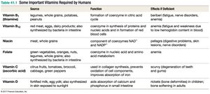
Important Vitamins Required by Humans
Vitamin | Source | Function | Effect if Deficient |
|---|---|---|---|
Vitamin B1 | Legumes, whole grains, potatoes | Coenzyme in citric acid cycle | Beriberi, fatigue, nerve disorders |
Vitamin B12 | Red meat, eggs, dairy | Synthesis of proteins and nucleic acids | Anemia, nervous system disorders |
Niacin | Meat, whole grains | Component of NAD+ and NADP+ | Pellagra, digestive problems, skin disorders |
Folate | Green vegetables, oranges, nuts | Coenzyme in nucleic acid and amino acid metabolism | Anemia |
Vitamin C | Citrus fruits, tomatoes, broccoli | Collagen synthesis, immune function | Scurvy, degeneration of teeth and gums |
Vitamin D | Sunlight, fortified milk, fish | Absorption of calcium and phosphorus | Rickets, bone deformities |
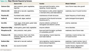
Major Minerals Required by Humans
Mineral | Source in Diet | Function | Effect if Deficient |
|---|---|---|---|
Calcium (Ca) | Dairy, green vegetables | Bone and tooth formation, nerve signaling | Loss of bone mass, slow growth |
Chloride (Cl) | Table salt, sea salt, vegetables | Fluid balance, digestion | Weakness, loss of muscle function |
Fluorine (F) | Fluoridated water, seafood | Maintenance of tooth enamel | High frequency of tooth decay |
Iodine (I) | Seafood, iodized salt | Thyroid hormone synthesis | Goiter |
Magnesium (Mg) | Whole grains, green vegetables | Enzyme cofactor, muscle function | Nerve disorders |
Phosphorus (P) | Dairy, meat, grains | Bone and tooth formation, nucleic acid synthesis | Weakness, loss of bone mass |
Potassium (K) | Dairy, meat, fruits | Nerve signaling, muscle response | Weakness, muscle cramps |
Sodium (Na) | Table salt, sea salt, seafood | Nerve signaling, muscle response | Weakness, muscle cramps |
Sulfur (S) | Any source of protein | Amino acid synthesis | Swollen tissues, impaired metabolism |
Digestion and Absorption
Digestion is essential for animals because they cannot directly acquire nutrients as individual molecules. Food must be broken down into small enough pieces for nutrients to be extracted, and waste materials eliminated.
Digestive Tract Designs
Digestive tracts are classified as incomplete or complete:
Incomplete digestive tracts: One opening for both ingestion and elimination (e.g., Hydra, planaria).
Complete digestive tracts: Two openings—mouth for ingestion, anus for elimination. Allows compartmentalization of processes and uninterrupted digestion.
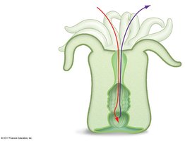
Advantages of Complete Digestive Tracts
Different processes can occur in specialized compartments.
One-way flow allows continuous ingestion and digestion.
Accessory organs (salivary glands, pancreas, liver, gallbladder) contribute enzymes and other products.
Digestive System Anatomy and Function
The mammalian digestive system consists of several organs, each with specialized functions:
Mouth: Mechanical and chemical processing; saliva digests carbohydrates.
Esophagus: Transports food via peristalsis.
Stomach: Mechanical and chemical processing; digests proteins.
Small intestine: Chemical processing and absorption; digests proteins, fats, carbohydrates; absorbs nutrients and water.
Large intestine: Absorbs water, forms feces; contains symbiotic bacteria.
Appendix: Contains immune tissue; harbors symbiotic bacteria.
Anus: Eliminates feces.
Liver: Secretes molecules aiding fat digestion; stores glucose and glycogen.
Gallbladder: Stores liver secretions; empties into small intestine.
Pancreas: Secretes enzymes and other materials into small intestine.
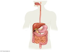
Overview of the Digestive Process
Digestion begins with chewing, followed by distinct chemical changes in each compartment:
Mouth: Enzymes in saliva break down carbohydrates and lipids.
Stomach: Acidic environment initiates protein digestion.
Small intestine: Completes chemical digestion and absorbs nutrients.
Large intestine: Absorbs water, forms feces.
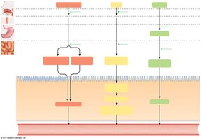
The Mouth and Esophagus
Salivary amylase hydrolyzes starch into maltose, a disaccharide. Lingual lipase from tongue cells begins lipid digestion. Mucus produced by salivary glands facilitates swallowing.
Peristalsis in the Esophagus
Food is moved to the stomach by peristalsis, a wave of muscle contractions triggered by swallowing.
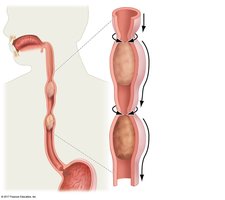
The Stomach
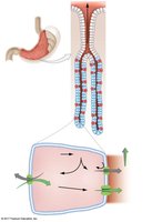
The stomach is a muscular pouch bracketed by sphincters. It churns food mechanically and partially digests proteins. The lumen is highly acidic due to hydrochloric acid (HCl).
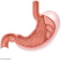
Protein Digestion in the Stomach
Pepsin: Enzyme responsible for protein digestion, produced as inactive pepsinogen by chief cells and activated by HCl.
Parietal cells: Source of HCl, which denatures proteins and activates pepsin.
Mucous cells: Secrete mucus to protect stomach lining.
Ulcers and Helicobacter pylori
Ulcers are eroded areas in the epithelium, often caused by infection with Helicobacter pylori, not just excess acid.
Ruminant Stomach
Ruminants (cattle, sheep, goats, deer, buffalo) have a four-chambered stomach specialized for digesting cellulose. The largest chamber, the rumen, houses symbiotic microbes that ferment cellulose.
The Small Intestine
Peristalsis moves material from the stomach to the small intestine, where digestion is completed and nutrients are absorbed. Protein digestion is finalized by pancreatic enzymes, yielding amino acids for transport in the bloodstream.
Digestive Enzymes in Mammals
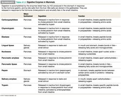
Enzyme | Where Synthesized | Regulation | Function |
|---|---|---|---|
Carboxypeptidase | Pancreas | Released in inactive form; activated by trypsin | Breaks peptide bonds in proteins |
Chymotrypsin | Pancreas | Released in inactive form; activated by trypsin | Breaks peptide bonds in proteins |
Elastase | Pancreas | Released in inactive form; activated by trypsin | Breaks peptide bonds in proteins |
Lingual lipase | Salivary glands | Released in active form | Breaks bonds in fats |
Pancreatic amylase | Pancreas | Released in active form | Breaks bonds in carbohydrates |
Pancreatic lipase | Pancreas | Released in active form | Breaks bonds in fats |
Pepsin | Stomach | Released in inactive form; activated by HCl | Breaks peptide bonds in proteins |
Salivary amylase | Salivary glands | Released in active form | Breaks bonds in carbohydrates |
Trypsin | Pancreas | Released in inactive form; activated by enteropeptidase | Breaks peptide bonds in proteins |
Water Absorption
Water is absorbed in the small intestine by osmosis, following the uptake of solutes. This process is crucial for reclaiming liquid secreted into the digestive tract.
The Large Intestine
By the time material reaches the large intestine, most water and nutrients have been absorbed. The colon forms feces by absorbing additional water and compacting waste. The rectum stores feces until elimination. Symbiotic bacteria in the colon digest cellulose and produce nutrients like vitamin K.
Appendix Function
The appendix, once considered vestigial, contains immune cells and serves as a safe haven for symbiotic bacteria, aiding recolonization after diarrhea.
Nutritional Homeostasis: Glucose Regulation
Glucose Homeostasis
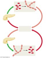
After digestion, nutrients enter the bloodstream and are delivered to cells. Glucose homeostasis is regulated by the hormones insulin and glucagon from the pancreas.
Insulin: Produced when blood glucose is high; stimulates glucose uptake and glycogen synthesis, lowering blood glucose.
Glucagon: Produced when blood glucose is low; stimulates glycogen breakdown and gluconeogenesis, raising blood glucose.
These hormones form a negative feedback system to maintain homeostasis.
Diabetes Mellitus
Diabetes mellitus is characterized by abnormally high blood glucose levels due to impaired insulin function.
Type 1 diabetes: Insufficient insulin synthesis (autoimmune destruction of insulin-producing cells).
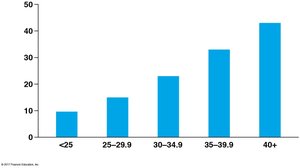
Type 2 diabetes: Insulin resistance (receptors do not function properly or are fewer); risk factors include obesity, high-sugar diets, lack of exercise, and genetics.
High blood glucose affects urine formation, increasing urine volume due to decreased water reabsorption.
Type 1 is treated with insulin injections and diet; type 2 is managed with diet, exercise, monitoring, and drugs to increase insulin responsiveness.

Summary Table: Diabetes Mellitus
Type | Cause | Treatment | Risk Factors |
|---|---|---|---|
Type 1 | Autoimmune destruction of insulin-producing cells | Insulin injections, diet | Genetic predisposition |
Type 2 | Insulin resistance | Diet, exercise, drugs | Obesity, high-sugar diet, lack of exercise, genetics |
Key Concepts and Applications
Animal nutrition involves ingestion, digestion, absorption, and elimination.
Essential nutrients must be obtained from the diet.
Digestive tract structure varies among animals, affecting function.
Accessory organs play critical roles in digestion.
Glucose homeostasis is vital for health; disruption leads to diabetes mellitus.
Example: The correlation between obesity and type 2 diabetes highlights the importance of diet and lifestyle in maintaining nutritional homeostasis. Additional info: Tables and diagrams have been expanded for clarity and completeness.
