 Back
Animal Responses to the Environment: Neurons, Synapses, and Sensory Systems
Study Guide - Smart Notes
 Tailored notes based on your materials, expanded with key definitions, examples, and context.
Animal Responses to the Environment
Introduction
Animals have evolved complex systems to detect, interpret, and respond to environmental stimuli. These adaptations involve specialized cells and organs that allow organisms to sense changes in their surroundings and coordinate appropriate responses. This section explores the structure and function of neurons, the organization of the nervous system, and the mechanisms underlying sensory perception and response.
Neuron Structure and Function
Types of Neurons
Sensory Neurons: Detect external or internal stimuli and transmit signals to the central nervous system (CNS).
Interneurons: Integrate information within the CNS and connect sensory and motor pathways.
Motor Neurons: Transmit signals from the CNS to effectors such as muscles or glands.
Organization of the Nervous System
Central Nervous System (CNS): Consists of the brain and spinal cord; responsible for processing and integrating information.
Peripheral Nervous System (PNS): Composed of nerves and ganglia outside the CNS; transmits signals between the CNS and the rest of the body.
Structure of Neurons and Glial Cells
Dendrites: Receive incoming signals from other neurons.
Axon: Conducts electrical impulses away from the cell body.
Axon Hillock: Region where action potentials are initiated.
Synapse: Junction between two neurons or between a neuron and an effector cell.
Glial Cells: Support, nourish, and protect neurons.
How Neurons Work: Action Potentials
Membrane Polarization
Resting Potential: The membrane potential of a neuron at rest, typically around -70 mV, maintained by the sodium-potassium pump and selective permeability of the membrane.
Depolarization: The inside of the cell becomes more positive due to the influx of Na+ ions.
Hyperpolarization: The inside of the cell becomes more negative, often due to the efflux of K+ ions.
Action Potential Steps
Depolarization: Voltage-gated Na+ channels open, allowing Na+ to enter the cell, making the inside more positive.
Repolarization/Hyperpolarization: Voltage-gated K+ channels open, K+ exits the cell, restoring negative charge inside.
Undershoot: Membrane potential temporarily becomes more negative than resting potential.
Resting Potential: Restored by the Na+/K+ pump.
All-or-None Response: An action potential only occurs if the membrane depolarizes above a threshold (about -55 mV). The magnitude of the action potential is independent of stimulus strength.
Conduction of Action Potentials
Action potentials are self-propagating and move in one direction along the axon.
Depolarization at one region triggers depolarization in the next region, similar to a domino effect.
Evolutionary Adaptations of Axons
Invertebrate axons are often large in diameter to increase conduction speed.
Vertebrate axons are typically myelinated, allowing for rapid signal transmission even with smaller diameters.
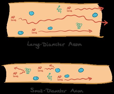
Saltatory Conduction: In myelinated axons, action potentials jump between Nodes of Ranvier, greatly increasing conduction speed.
Synapses and Neurotransmitters
Types of Synapses
Chemical Synapses: Use neurotransmitters to transmit signals across a synaptic cleft. Most common, slower, but allow for flexibility and adaptation (important for learning and memory).
Electrical Synapses: Allow direct flow of ions through gap junctions; very fast, important for rapid reflexes.
Chemical Synaptic Signaling Steps
An action potential arrives at the presynaptic terminal, depolarizing the membrane.
Voltage-gated Ca2+ channels open, and Ca2+ enters the terminal.
Synaptic vesicles containing neurotransmitters fuse with the presynaptic membrane.
Neurotransmitters are released into the synaptic cleft and diffuse across to the postsynaptic membrane.
Neurotransmitters bind to receptors, triggering a response in the postsynaptic cell.
Neurotransmitters are either degraded or reabsorbed by the presynaptic neuron.
Sensory Receptors and Perception
Types of Sensory Receptors
Mechanoreceptors: Detect mechanical forces such as pressure, touch, stretch, motion, and sound.
Electromagnetic Receptors: Detect light, electricity, and magnetism.
Thermoreceptors: Detect changes in temperature.
Pain Receptors (Nociceptors): Detect tissue damage or potentially harmful stimuli.
Chemoreceptors: Detect chemical changes, such as glucose, oxygen, carbon dioxide, and amino acids.
Example: Spicy foods activate the same receptors as hot temperatures, while menthol activates cold receptors.
Mechanoreceptors and Hearing
Sensory organs for hearing and balance are closely associated in vertebrates.
The cochlea is involved in hearing, while semicircular canals function in equilibrium.
Sound waves cause the tympanic membrane (eardrum) to vibrate, which is transmitted via middle ear bones to the cochlea, creating pressure waves in cochlear fluid.
These waves bend hair cells, opening ion channels and sending signals to the auditory nerve and brain.
Photoreceptors and Vision
Photoreceptors contain light-absorbing pigment molecules.
Vertebrates use single-lens eyes; light enters through the pupil, and the iris adjusts light entry.
The lens changes shape to focus light on the retina, which contains rods (for night vision) and cones (for color vision).
Signals are relayed via bipolar and ganglion cells to the optic nerve and visual cortex.
Optic Disc: The region where the optic nerve exits the eye; lacks rods and cones, creating a blind spot.
Summary Table: Types of Sensory Receptors
Receptor Type | Stimulus Detected | Examples |
|---|---|---|
Mechanoreceptors | Mechanical forces | Touch, hearing, balance |
Electromagnetic receptors | Light, electricity, magnetism | Vision (photoreceptors) |
Thermoreceptors | Temperature changes | Heat, cold |
Pain receptors (nociceptors) | Tissue damage | Pain sensation |
Chemoreceptors | Chemical changes | Smell, taste, blood chemistry |
Key Equations
Nernst Equation (for equilibrium potential):
Resting Membrane Potential (Goldman-Hodgkin-Katz Equation):
Additional info: This guide expands on the provided notes with definitions, examples, and equations relevant to the structure and function of neurons, synapses, and sensory systems, as covered in college-level biology courses.
