 Back
BackAnimal Sensory and Motor Mechanisms: Sensory Pathways, Muscle Function, and Skeletal Systems
Study Guide - Smart Notes
 Tailored notes based on your materials, expanded with key definitions, examples, and context.
Animal Sensory and Motor Mechanisms
Overview
This study guide covers the structure and function of animal sensory systems, muscle contraction, and skeletal systems, focusing on the mechanisms by which animals detect and respond to their environment. It is organized according to the major concepts in animal physiology and is suitable for college-level biology students.
Concept 50.1: Sensory Receptors and Sensory Pathways
Sensory Pathway Functions
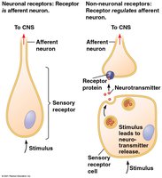
Sensory Reception: Detection of stimuli by sensory receptors (cells or organs that interact with internal or external stimuli).
Transduction: Conversion of stimulus energy into a change in membrane potential (receptor potential) in the sensory receptor.
Transmission: Sensory information travels as action potentials to the central nervous system (CNS).
Perception: The brain interprets the incoming signals, constructing a representation of the stimulus.
Sensory Reception and Transduction
Sensory receptors can be either neurons or specialized non-neuronal cells. They initiate the sensory pathway by detecting stimuli and converting them into electrical signals.
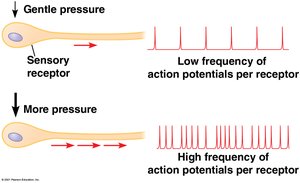
Key Point: Receptor potentials are graded, meaning their magnitude varies with stimulus strength.
Transmission
The intensity of a stimulus is encoded by the frequency of action potentials generated by the sensory neuron.
Perception
Perceptions are created by the brain based on the neural pathway activated.
Different sensory modalities (e.g., vision, hearing) are distinguished by the CNS based on the origin of the action potentials.
Amplification and Adaptation
Amplification: Strengthening of a sensory signal during transduction.
Sensory Adaptation: Decreased responsiveness to a continued stimulus.
Types of Sensory Receptors
Mechanoreceptors: Detect mechanical energy (touch, pressure, vibration, stretch).
Chemoreceptors: Detect chemical stimuli (solute concentration, specific molecules).
Electromagnetic Receptors: Detect light, electricity, magnetism.
Thermoreceptors: Detect temperature changes.
Pain Receptors (Nociceptors): Detect harmful conditions (extreme pressure, temperature, chemicals).
Mechanoreceptors and Other Sensory Receptors
Mechanoreceptors
Mechanoreceptors are responsible for sensing physical deformation caused by mechanical forces. In mammals, these are often dendrites of sensory neurons located in the skin and other tissues.

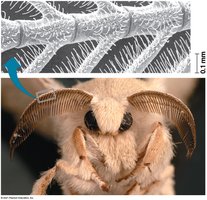
Chemoreceptors
Chemoreceptors can detect either the total solute concentration or specific molecules. For example, the antennae of male silkworm moths have highly sensitive chemoreceptors for pheromones.
Electromagnetic Receptors
These receptors detect electromagnetic energy. Some animals, such as the platypus, use electroreceptors to locate prey, while others use magnetic fields for navigation.
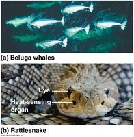
Thermoreceptors
Thermoreceptors detect heat and cold. For example, rattlesnakes have specialized organs to sense infrared radiation from warm-blooded prey.
Pain Receptors
Pain receptors (nociceptors) detect harmful stimuli, such as extreme pressure, temperature, or chemicals released from damaged tissues.
Concept 50.2: Hearing and Equilibrium
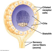
Hearing and Equilibrium in Invertebrates
Many invertebrates use statocysts, which contain mechanoreceptors and statoliths, to sense gravity and maintain equilibrium.
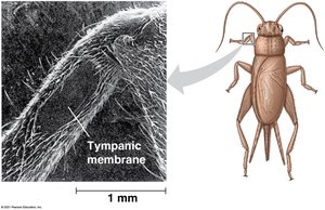
Some insects detect sound using body hairs or tympanic membranes.
Hearing and Equilibrium in Mammals
The mammalian ear is divided into three regions: outer, middle, and inner ear. The ear transduces sound waves into nerve impulses and also detects equilibrium (balance).
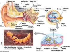
Outer Ear: Captures and funnels sound waves.
Middle Ear: Contains ossicles (malleus, incus, stapes) that amplify vibrations.
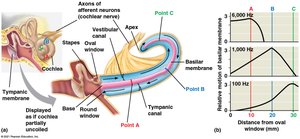
Inner Ear: Contains the cochlea (hearing) and vestibular system (equilibrium).
Mechanism of Hearing
Sound waves cause vibrations in the tympanic membrane, which are transmitted and amplified by the ossicles to the cochlea. Hair cells in the cochlea transduce these vibrations into electrical signals.

Action Potentials in Hearing
Bending of hair cell stereocilia opens mechanically gated K+ channels, leading to depolarization and generation of action potentials.

Equilibrium
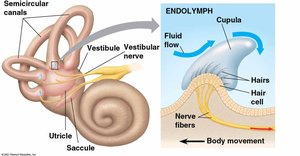
The utricle, saccule, and semicircular canals in the inner ear detect linear and angular movements, allowing the brain to perceive balance and orientation.
Hearing and Equilibrium in Other Vertebrates
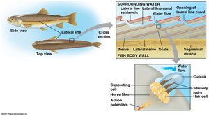
Fishes and aquatic amphibians have a lateral line system containing mechanoreceptors that detect water movement.
Concept 50.3: Visual Receptors and Vision
Evolution of Visual Perception
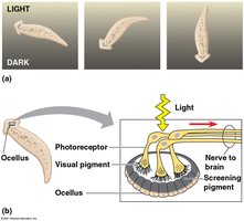
All visual systems rely on photoreceptors containing light-absorbing pigments. Planarians have simple eyespots (ocelli) for detecting light direction.
Compound Eyes
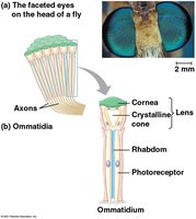
Insects and crustaceans have compound eyes made of many ommatidia, each functioning as a separate light detector. Compound eyes are excellent for detecting movement and color.
Single-Lens Eyes
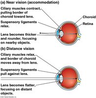
Vertebrates and some invertebrates have single-lens eyes, which focus light onto a retina using a lens and iris.
The Vertebrate Visual System
The vertebrate eye consists of external and internal structures that focus and process light. The retina contains rods (sensitive to low light) and cones (color vision).

How We See
Light is focused onto the retina, where photoreceptors (rods and cones) transduce it into electrical signals. These signals are processed by bipolar and ganglion cells, then sent to the brain via the optic nerve.
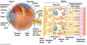
Sensory Transduction in the Eye
In darkness, Na+ channels are open in photoreceptors.
Light activates rhodopsin, which triggers a cascade leading to closure of Na+ channels and hyperpolarization.
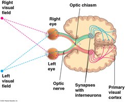
Processing Visual Information
Axons from ganglion cells form the optic nerve, which crosses at the optic chiasm. Visual information from each visual field is processed in the opposite hemisphere of the brain.
Color Vision
Humans have three types of cones (red, green, blue), each with a different photopsin pigment. Most mammals have less acute color vision than primates.
The Visual Field
The fovea is the center of the visual field, densely packed with cones for sharp vision.
Concept 50.4: Taste and Smell
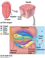
Taste (Gustation) in Mammals
There are five basic tastes: sweet, sour, salty, bitter, and umami. Taste receptor cells are organized into taste buds, mainly on the tongue.
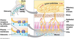
Smell (Olfaction) in Humans
Olfactory receptor neurons in the nasal cavity detect odorant molecules. Each neuron expresses one type of olfactory receptor gene, and signals are sent to the olfactory bulb in the brain.
Concept 50.5: Muscle Function and Contraction
Structure of Skeletal Muscle
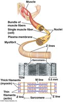
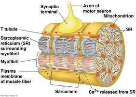
Skeletal muscle is composed of bundles of muscle fibers, each containing myofibrils made of repeating units called sarcomeres. Sarcomeres are the functional units of contraction.
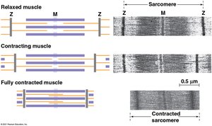
The Sliding-Filament Model
Muscle contraction occurs as thin (actin) and thick (myosin) filaments slide past each other, shortening the sarcomere.
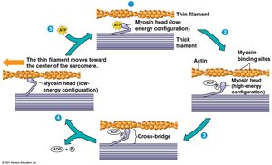
The Cross-Bridge Cycle
Myosin heads bind to actin, perform a power stroke, release upon ATP binding, and reset for another cycle.
Role of Calcium and Regulatory Proteins
Tropomyosin and the troponin complex block myosin-binding sites on actin when the muscle is relaxed.
Ca2+ binding to troponin exposes these sites, allowing contraction.
Signal Transduction in Muscle Fibers
Action potentials from motor neurons trigger Ca2+ release from the sarcoplasmic reticulum, initiating contraction.
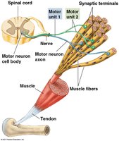
Nervous Control of Muscle Tension
Muscle contraction strength is regulated by the number of fibers activated and the rate of stimulation. A motor unit consists of a motor neuron and all the muscle fibers it controls.
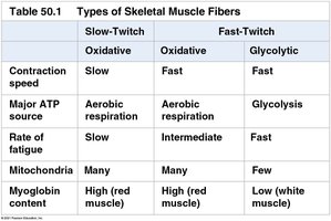
Types of Skeletal Muscle Fibers
Muscle fibers are classified by contraction speed and ATP source:
Slow-Twitch (Oxidative) | Fast-Twitch (Oxidative) | Fast-Twitch (Glycolytic) | |
|---|---|---|---|
Contraction speed | Slow | Fast | Fast |
Major ATP source | Aerobic respiration | Aerobic respiration | Glycolysis |
Rate of fatigue | Slow | Intermediate | Fast |
Mitochondria | Many | Many | Few |
Myoglobin content | High (red muscle) | High (red muscle) | Low (white muscle) |
Concept 50.6: Skeletal Systems and Locomotion
Types of Skeletal Systems
Hydrostatic Skeletons: Fluid-filled compartments provide support (e.g., annelids).
Exoskeletons: Hard external coverings (e.g., arthropods, molluscs).
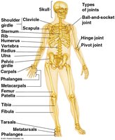
Endoskeletons: Internal skeletons (e.g., vertebrates) made of bone and/or cartilage.
Types of Locomotion
Land: Walking, running, hopping, crawling (overcoming gravity).
Swimming: Streamlined bodies reduce friction in water.
Flying: Wings generate lift to overcome gravity; adaptations reduce body mass.
Additional info: This guide integrates and expands upon the provided lecture content, ensuring all major concepts from animal sensory and motor systems are covered for exam preparation.
