Back
Animal Tissues and Organs: Structure, Function, and Organization
Study Guide - Smart Notes
 Tailored notes based on your materials, expanded with key definitions, examples, and context.
Hierarchical Organization of Animal Structure
Levels of Biological Organization
The animal body is organized into a hierarchy of structural levels, each with increasing complexity and specialization. Understanding these levels is fundamental to the study of animal biology and physiology.
Cells: The basic unit of structure and function in animals, each with specialized roles.
Tissues: Groups of similar cells that perform a common function.
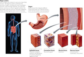
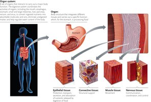
Organs: Structures composed of multiple tissue types working together for specific tasks.
Organ Systems: Groups of organs that coordinate to carry out major physiological processes.
Animal Cell Connections
Types of Intercellular Junctions
Animal cells are connected by specialized junctions that facilitate communication, adhesion, and barrier formation.
Gap Junctions: Protein complexes that form channels between cells, allowing ions and small molecules to pass. Analogous to plasmodesmata in plants.
Tight Junctions: Fused membrane proteins that prevent leakage of substances between cells, maintaining compartmentalization.
Anchoring Junctions: Filamentous structures that tie cytoskeletons of adjacent cells together, providing mechanical stability.
Main Types of Animal Tissues
Overview of the Four Tissue Types
Animal tissues are classified into four main types, each with distinct structures and functions essential for organismal survival.
Epithelial Tissue: Covers body surfaces and lines cavities; involved in protection, absorption, and secretion.
Connective Tissue: Provides support, binds tissues, and stores energy; includes bone, blood, and adipose tissue.
Muscle Tissue: Specialized for contraction and movement.
Nervous Tissue: Conducts electrical impulses and processes information.
Epithelial Tissue
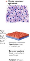
Simple Squamous Epithelium
Simple squamous epithelium consists of a single layer of flattened cells, facilitating rapid diffusion and filtration.
Function: Diffusion and filtration of gases and small molecules.
Structure: Single, thin layer of flat cells.
Locations: Air sacs of lungs, lining of blood vessels, and portions of the gastrointestinal tract.
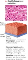
Stratified Squamous Epithelium
Stratified squamous epithelium is composed of multiple layers of cells, providing protection against abrasion and mechanical stress.
Function: Protection from abrasion and pathogens.
Structure: Several layers of flattened cells, with cells at different stages of life and function.
Locations: Skin, mouth, esophagus, and other surfaces subject to friction.
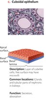
Cuboidal Epithelium
Cuboidal epithelium consists of cube-shaped cells specialized for secretion and absorption.
Function: Secretion and absorption of substances.
Structure: Single layer of cuboidal cells, often with microvilli to increase surface area.
Locations: Glands and ducts, such as those in the kidneys and endocrine glands.
Columnar Epithelium
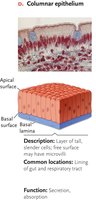
Columnar epithelium is made up of tall, column-like cells, often involved in absorption and secretion.
Function: Absorption and secretion, especially of mucus and enzymes.
Structure: Single layer of elongated cells, sometimes with microvilli or cilia.
Locations: Lining of the gastrointestinal tract and respiratory passages.
Epithelial Glands
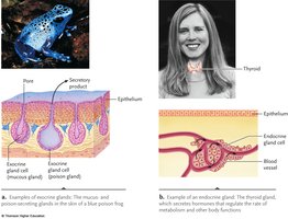
Glands are specialized epithelial structures that secrete substances necessary for bodily functions.
Exocrine Glands: Secrete products through ducts to external or internal surfaces (e.g., sweat, oil, digestive glands).
Endocrine Glands: Ductless glands that release hormones directly into the bloodstream or extracellular fluid (e.g., thyroid, pancreas).
Connective Tissue
Types of Connective Tissue
Connective tissues support, bind, and protect other tissues and organs. They are characterized by an abundant extracellular matrix.
Loose Connective Tissue: Provides support, elasticity, and diffusion; found under epithelial layers.
Fibrous Connective Tissue: Dense with collagen fibers, providing strength and elasticity; found in tendons and ligaments.
Cartilage: Flexible, impact-resistant tissue found in joints, airways, and embryonic skeletons.
Bone: Rigid tissue for support, movement, and protection; contains osteocytes in a mineralized matrix.
Adipose Tissue: Stores fat for energy, insulation, and protection.
Blood: Fluid tissue for transport of gases, nutrients, and waste; contains erythrocytes, leukocytes, and platelets in plasma.
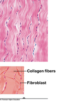
Fibrous Connective Tissue
Function: Strength and elasticity.
Structure: Parallel collagen and elastin fibers with fibroblasts.
Locations: Tendons and ligaments.
Cartilage
Function: Support and low-friction surfaces for joints.
Structure: Chondrocytes in a pliable matrix of collagen.
Locations: Joints, airways, embryonic skeletons.
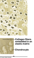
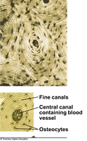
Bone
Function: Movement, support, and protection.
Structure: Osteocytes in a matrix of collagen and minerals (calcium, magnesium).
Locations: Vertebrate skeletons.
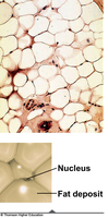
Adipose Tissue
Function: Insulation, protection, and energy storage.
Structure: Large, tightly packed adipocytes with minimal extracellular matrix.
Locations: Under skin, around organs.
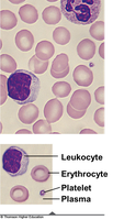
Blood
Function: Transport of substances throughout the body.
Structure: Cells (erythrocytes, leukocytes, platelets) suspended in plasma.
Locations: Circulatory system.
Muscle Tissue
Types of Muscle Tissue
Muscle tissue is specialized for contraction, enabling movement and force generation in the body. There are three main types:
Skeletal Muscle: Voluntary movement, posture; long, multinucleated fibers attached to bones.
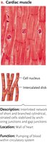
Cardiac Muscle: Involuntary contraction of the heart; branched cells with intercalated disks for electrical connectivity.
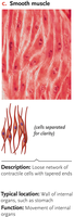
Smooth Muscle: Involuntary movement of internal organs; spindle-shaped cells in walls of digestive tract, blood vessels, etc.
Cardiac Muscle
Function: Pumping blood through the circulatory system.
Structure: Branched, interconnected cells with intercalated disks.
Location: Heart wall.
Smooth Muscle
Function: Movement of internal organs, sustained contractions.
Structure: Spindle-shaped, non-striated cells.
Locations: Walls of digestive tract, blood vessels, sphincters, ducts.
Nervous Tissue
Structure and Function
Nervous tissue is responsible for communication and control within the body, using electrical and chemical signals.
Neurons: Specialized cells that transmit nerve impulses between tissues, organs, and systems.
Glial Cells: Support neurons, provide insulation, and supply nutrients.
Integration: Tissues, Organs, and Organ Systems
From Cells to Organ Systems
Cells form tissues, which combine to create organs. Organs work together in organ systems to perform complex physiological functions necessary for survival.
Example: The digestive system includes organs such as the esophagus, stomach, and colon, each composed of multiple tissue types (epithelial, connective, muscle, nervous) to process and absorb nutrients.