 Back
L 3.1: Bacteria: Diversity, Structure, and Identification – Study Notes
Study Guide - Smart Notes
 Tailored notes based on your materials, expanded with key definitions, examples, and context.
L 3.1
Bacterial Diversity and Taxonomy
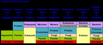
Historical Evolution of Classification
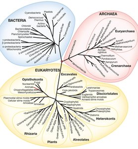
The classification of living organisms has evolved from a two-kingdom system to a six-kingdom and three-domain system, reflecting advances in molecular biology and genetics. Modern taxonomy recognizes Bacteria, Archaea, and Eukarya as the three domains of life, with further subdivisions into kingdoms based on genetic and phenotypic characteristics.
Early Systems: Linnaeus (two kingdoms: Plantae and Animalia), Haeckel (added Protista), and later systems introduced Monera (prokaryotes) and Fungi.
Modern Systems: The three-domain system (Bacteria, Archaea, Eukarya) is based on molecular data, especially 16S rRNA gene sequencing.
Domain Archaea
Characteristics and Habitats
Archaea are prokaryotic microorganisms distinct from bacteria, often inhabiting extreme environments but also found in more common habitats. They possess unique membrane lipids and genetic sequences.
Extreme Environments: High temperature (hydrothermal vents), high salinity (salt lakes), acidic or alkaline waters, and anoxic environments (marshes, digestive tracts).
Metabolic Diversity: Some archaea are methanogens, halophiles, or thermophiles.


Domain Bacteria
Bacterial Morphology
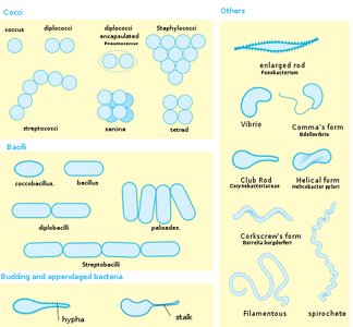
Bacteria exhibit a variety of shapes and arrangements, which are important for identification and classification.
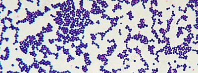
Cocci: Spherical bacteria (e.g., Streptococcus, Staphylococcus).
Bacilli: Rod-shaped bacteria (e.g., Escherichia, Bacillus).
Spirilla/Spirochetes: Spiral-shaped bacteria (e.g., Spirillum, Treponema).
Other Forms: Vibrio (comma-shaped), filamentous, and appendaged bacteria.
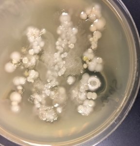
Bacterial Colonies
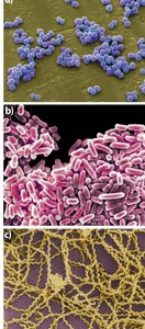
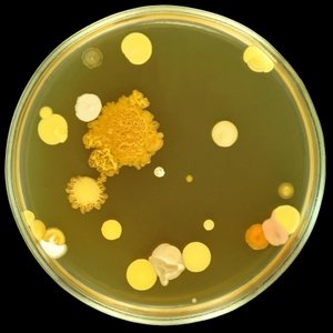
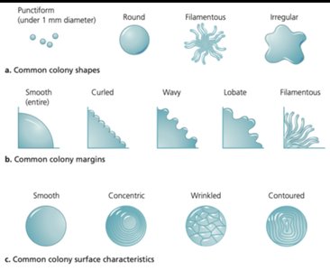
Bacterial colonies are visible masses of microbial cells arising from a single cell or group. Colony morphology (shape, margin, surface) aids in identification.
Colony Shape: Round, filamentous, irregular, etc.
Colony Margin: Smooth, wavy, lobate, filamentous.
Surface Characteristics: Smooth, concentric, wrinkled, contoured.
Gram Stain and Cell Wall Structure
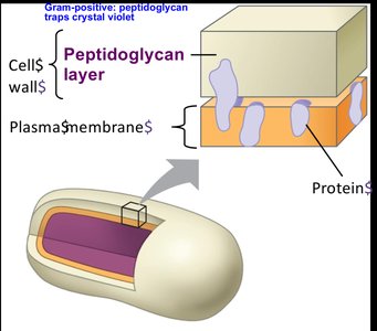
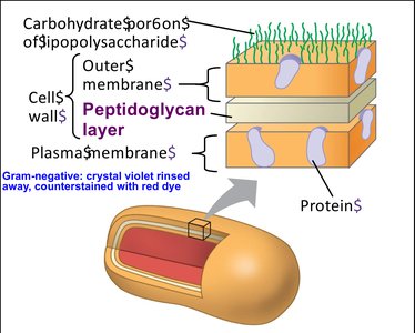
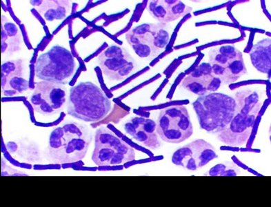
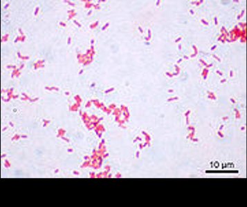
The Gram stain is a differential staining technique that distinguishes bacteria based on cell wall composition. It is a fundamental tool in microbiology for bacterial identification.
Gram-Positive Bacteria: Thick peptidoglycan layer, stains purple.
Gram-Negative Bacteria: Thin peptidoglycan layer, outer membrane, stains pink/red.
Gram Stain Procedure: 1. Heat-fix smear, 2. Crystal violet, 3. Iodine, 4. Decolorize, 5. Safranin counterstain.
Acid-Fast Stain
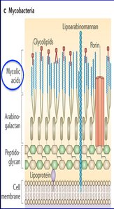
The acid-fast stain differentiates bacteria with mycolic acids in their cell walls, such as Mycobacterium species (e.g., tuberculosis, leprosy).
Mycolic Acids: Waxy, hydrophobic cell wall components that retain primary stain even after acid-alcohol decolorization.
Clinical Importance: Used to identify Mycobacterium tuberculosis and M. leprae.
Bacterial Phyla
Major Bacterial Groups
Bacteria are classified into several phyla based on genetic and phenotypic characteristics. Key phyla include:
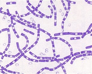
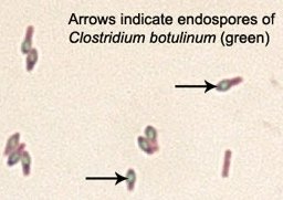
Firmicutes: Low GC Gram-positive bacteria (e.g., Bacillus, Clostridium, Staphylococcus).
Actinobacteria: High GC Gram-positive bacteria (e.g., Streptomyces, Mycobacterium).
Proteobacteria: Large group of Gram-negative bacteria (e.g., Escherichia, Salmonella, Pseudomonas).
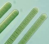
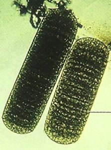
Cyanobacteria: Photosynthetic bacteria, some fix nitrogen (e.g., Anabaena, Oscillatoria).
Other Phyla: Spirochaetes (Treponema), Chlamydiae (Chlamydia), Bacteroidetes (Bacteroides).
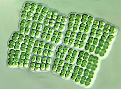
Cyanobacteria
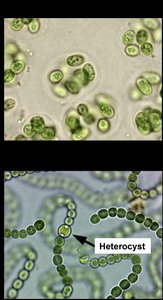
Cyanobacteria are oxygenic photosynthetic bacteria, important for nitrogen fixation and primary production in aquatic environments.
Heterocysts: Specialized cells for nitrogen fixation.
Akinetes: Thick-walled dormant cells for survival.
Examples: Anabaena, Oscillatoria, Merismopedia, Nostoc, Gleocapsa.
Bacterial Identification Techniques
Staining Methods
Gram Stain: Differentiates Gram-positive and Gram-negative bacteria.
Acid-Fast Stain: Identifies mycobacteria.
Capsule Stain: Visualizes gelatinous capsules, important for virulence.
Giemsa Stain: Used for blood parasites and some bacteria.
Biochemical Testing
Bacteria are identified by their metabolic capabilities, such as fermentation of sugars or utilization of specific substrates. Clinical labs use panels of biochemical tests (e.g., Enterotube).
Culturing and Colony Morphology
Bacteria are grown on various media to observe colony morphology, pigmentation, and hemolysis patterns, aiding in identification.
Antibiotics and Bacterial Control
Definition and Mechanism
An antibiotic is a chemical produced by a microorganism that inhibits or kills other microorganisms. Antibiotics are selective, targeting specific groups of bacteria or fungi, and are used in medicine to treat infections.
Zone of Inhibition: The area around an antibiotic disk where bacteria do not grow, used to measure antibiotic effectiveness.
Therapeutic Index: The ratio of the toxic dose to the effective dose, indicating safety for human use.
Laboratory Safety and Aseptic Technique
Best Practices
Proper aseptic technique is essential to prevent contamination of cultures and protect laboratory personnel and the environment.
Wear protective clothing (lab coat, gloves).
Disinfect work surfaces before and after use.
Wash hands before and after handling cultures.
Flame instruments before and after use.
Dispose of contaminated materials properly.
Summary Table: Gram-Positive vs. Gram-Negative Bacteria
Feature | Gram-Positive | Gram-Negative |
|---|---|---|
Peptidoglycan Layer | Thick | Thin |
Outer Membrane | Absent | Present |
Stain Color | Purple | Pink/Red |
Teichoic Acids | Present | Absent |
Examples | Staphylococcus, Bacillus | Escherichia, Pseudomonas |
Additional info:
Metagenomics allows the study of microbial diversity in environments where most microbes cannot be cultured.
Endospores are highly resistant structures formed by some Gram-positive bacteria (e.g., Bacillus, Clostridium).
