 Back
BackBacterial Cell Structure, Staining, and Laboratory Techniques
Study Guide - Smart Notes
 Tailored notes based on your materials, expanded with key definitions, examples, and context.
Bacterial Cell Structure and Morphology
Cell Shape and Arrangement
Bacteria exhibit a variety of shapes and arrangements, which are important for identification and classification. Common shapes include cocci (spherical), bacilli (rod-shaped), and spirilla (spiral-shaped). The arrangement of cells, such as clusters, chains, or pairs, is determined by the pattern of cell division and whether cells remain attached after division.
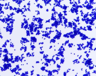
Cocci: Spherical bacteria, often found in clusters (e.g., Staphylococcus) or chains (e.g., Streptococcus).
Bacilli: Rod-shaped bacteria, which may occur singly or in chains.
Spirilla: Spiral-shaped bacteria, less common but distinctive.
Arrangement: Determined by cell division and adhesion; can be diagnostic.
Example: Staphylococcus epidermidis forms grape-like clusters of spheres.
Laboratory Preparation and Staining Techniques
Wet Mount and Thin Smear Preparation
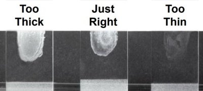
Wet mounts and thin smears are essential for observing bacterial morphology under a microscope. Proper technique ensures cells are spaced and fixed, preserving their native shape and arrangement.
Wet Mount: Used for live observation; bacteria appear transparent and out of focus.
Thin Smear: Created by spreading a small amount of culture on a slide, air drying, and heat fixing to preserve cell structure.
Heat Fixation: Melts proteins, anchoring cells to the slide.
Example: A correctly prepared thin smear allows clear observation of cell shape and arrangement.
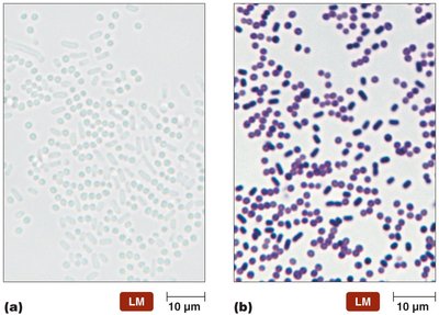
Simple Staining
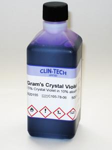
Simple stains use a single dye to increase contrast, making bacterial cells more visible. Crystal violet is commonly used for this purpose.
Procedure: Flood smear with crystal violet, wait 1 minute, rinse gently, and pat dry with bibulous paper.
Result: Cells appear purple, allowing for easy identification of morphology.
Gram Staining
Gram staining differentiates bacteria based on cell wall structure, classifying them as Gram positive or Gram negative. This is a fundamental technique in microbiology.
Gram Positive: Thick peptidoglycan layer traps crystal violet, cells appear purple.
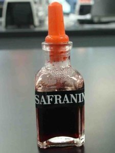
Gram Negative: Thin peptidoglycan layer and outer membrane; crystal violet is washed away, cells appear pink after counterstaining with safranin.
Steps:
Flood smear with crystal violet (primary stain).
Apply iodine (mordant).
Decolorize with 95% ethyl alcohol.
Counterstain with safranin.

Gram Stain Results
Gram stain results are interpreted based on color and morphology. Gram positive cells retain the purple color, while Gram negative cells appear pink.
Gram Positive Example: Staphylococcus epidermidis (purple, clustered cocci).
Gram Negative Example: Escherichia coli (pink, rod-shaped).
Bacterial Cell Wall Structure
Gram Positive Cell Wall
Gram positive bacteria have a thick peptidoglycan layer, which constitutes about 90% of the cell wall. Teichoic acids provide stability, and lipoteichoic acids anchor the peptidoglycan to the cell membrane.
Peptidoglycan: Acts as a sponge, trapping crystal violet dye.
Teichoic Acids: Contribute to cell wall stability.
Lipoteichoic Acids: Anchor peptidoglycan to membrane lipids.
Gram Negative Cell Wall
Gram negative bacteria have a thin peptidoglycan layer (about 10% of cell wall) and an outer lipid bilayer membrane containing phospholipids, proteins, and lipopolysaccharides. The outer membrane blocks crystal violet from reaching the peptidoglycan.
Outer Membrane: Contains lipopolysaccharides, provides protection.
Peptidoglycan: Thin layer, less dye retention.
Permeability: Outer membrane restricts entry of certain substances.
Microscopy Techniques
Light Microscopy: Simple vs. Compound Microscopes
Microscopes are essential tools for observing bacteria. Simple microscopes use a single lens, while compound microscopes use multiple lenses for greater magnification and resolution.
Simple Microscope: Single lens, limited magnification.
Compound Microscope: Objective and ocular lenses, higher magnification.
Total Magnification Formula:
Example: High power objective (40x) and ocular (10x):
Bacterial Culture and Aseptic Technique
Culture Media Types
Bacteria are grown in laboratory settings using various media types. Proper aseptic technique prevents contamination.
Liquid Broth: Growth indicated by turbidity.
Agar Plate: Growth indicated by colonies or lawn.
Agar Slant: Growth indicated by colonies or lawn on slant surface.
Aseptic Technique: Sterilization of equipment and media to prevent contamination.
Bacterial Transfer Techniques
Bacteria are transferred between media using sterilized inoculating loops or needles. Pure cultures contain one species; mixed cultures contain multiple species and require streaking techniques for isolation.
Inoculating Loop/Needle: Sterilized before and after transfer.
Three Way Streak Plate: Used to isolate individual colonies from mixed cultures.
Colony Morphology
Physical Characteristics of Colonies
Colony morphology is used to identify bacteria. Characteristics include size, elevation, color, surface, edge, and odor.
Size: Used for comparison between species.
Elevation: Flat, raised, convex, umbonate, crateriform, plateau.
Color: Pigment production varies by species.
Surface: Dry, shiny, powdery, or glistening depending on glycocalyx.
Edge: Entire, undulate, erose, spreading, rhizoid.
Odor: Some species produce distinctive odors (e.g., Pseudomonas).
Example: Proteus mirabilis colonies are smooth, grey, large, and contain circular rings.
Summary Table: Gram Positive vs. Gram Negative Cell Walls
Feature | Gram Positive | Gram Negative |
|---|---|---|
Peptidoglycan Thickness | Thick (90%) | Thin (10%) |
Teichoic Acids | Present | Absent |
Outer Membrane | Absent | Present |
Crystal Violet Retention | High (purple) | Low (pink after safranin) |
Example Species | Staphylococcus epidermidis | Escherichia coli |
Additional info: Colony morphology can be influenced by environmental conditions and media type, and is determined by genetics, pigment production, cell wall type, motility, and metabolism.
