 Back
BackBacterial Cell Structure, Staining, and Laboratory Techniques
Study Guide - Smart Notes
 Tailored notes based on your materials, expanded with key definitions, examples, and context.
Bacterial Cell Structure and Morphology
Introduction to Bacteria
Bacteria are single-celled prokaryotic organisms that exhibit a variety of shapes and arrangements. Observing bacteria under the microscope provides essential information about their morphology, which is critical for identification and classification.
Shape: Common bacterial shapes include cocci (spherical), bacilli (rod-shaped), and spirilla (spiral).
Arrangement: Bacteria may occur singly, in pairs, chains, clusters, or other groupings depending on the species and mode of division.
Observation: Wet mounts and staining techniques are used to visualize bacteria, as they are typically transparent and difficult to see without contrast enhancement.
Microscopy and Slide Preparation
Wet Mounts and Smear Preparation
Preparing a proper bacterial smear is essential for reliable staining and observation. The smear must be thin and evenly distributed to preserve the native shape and arrangement of cells.
Broth Culture: Transfer a loopful of liquid culture to a slide.
Agar Slant/Plate: Mix a small amount of colony with a drop of distilled water on the slide.
Air Dry: Allow the smear to air dry to maintain cell structure.
Heat Fixation: Pass the slide through a flame to fix cells to the slide by melting proteins.
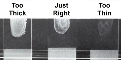
Note: Smears that are too thick result in clumping, while smears that are too thin may not show enough cells for observation.
Staining Techniques
Simple Stain
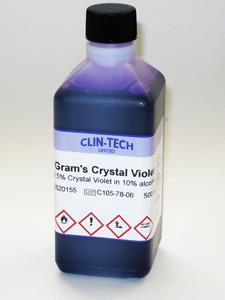
Simple staining uses a single dye to increase contrast, making bacterial cells visible under the microscope. Crystal violet is a common dye used for this purpose.
Flood the smear with crystal violet for 1 minute.
Rinse gently with water above the smear to avoid washing off cells.
Pat dry with bibulous paper.
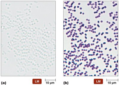
Result: Bacterial cells appear colored, allowing for observation of shape and arrangement.
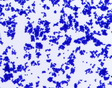
Gram Stain
The Gram stain is a differential staining technique that distinguishes bacteria based on cell wall structure. It is one of the most important techniques in microbiology for bacterial classification.
Flood the smear with crystal violet (primary stain) for 1 minute.
Rinse with water.
Add iodine (mordant) to form a crystal violet-iodine complex.
Decolorize with 95% ethyl alcohol for 10–20 seconds.
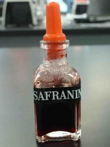
Counterstain with safranin for 1 minute.
Rinse and pat dry.
Results:
Gram-positive bacteria: Retain crystal violet and appear purple due to a thick peptidoglycan layer.
Gram-negative bacteria: Lose crystal violet during decolorization and take up safranin, appearing pink/red due to a thin peptidoglycan layer and an outer membrane.
Bacterial Cell Wall Structure
Gram-Positive Cell Wall
Gram-positive bacteria have a thick peptidoglycan layer, which retains the crystal violet dye during Gram staining. Teichoic acids are present, providing structural stability and anchoring the peptidoglycan to the cell membrane.
Peptidoglycan makes up about 90% of the cell wall.
Teichoic and lipoteichoic acids are important components.
Gram-Negative Cell Wall
Gram-negative bacteria have a thin peptidoglycan layer (about 10% of the cell wall) and an outer lipid bilayer membrane containing phospholipids, proteins, and lipopolysaccharides. The outer membrane prevents crystal violet from being trapped, so these cells are decolorized and counterstained with safranin.
Outer membrane contains lipopolysaccharides (LPS), which are important for pathogenicity and immune response.
Thin peptidoglycan layer is located between the outer and inner membranes.

Laboratory Culture Techniques
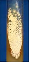
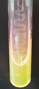
Media Types and Growth
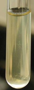
Bacteria can be cultured in liquid (broth) or on solid (agar) media. The choice of media and technique depends on the experimental goal, such as isolating pure colonies or preparing cells for staining.
Broth: Growth indicated by turbidity (cloudiness).
Agar Plate: Growth indicated by visible colonies or a lawn.
Agar Slant: Used for storage and observation of colony morphology.
Aseptic Technique
Aseptic techniques are essential to prevent contamination of cultures and ensure reliable results. This includes sterilizing equipment, media, and using proper transfer methods.
Use of inoculating loops and needles, sterilized by flame or heat.
Autoclaving media at 121°C and 15 psi for 15 minutes to eliminate microbes.
Proper handling of test tubes and petri dishes to avoid introducing contaminants.
Colony Morphology
Colony morphology refers to the physical characteristics of bacterial colonies, which can aid in identification. Features include size, elevation, color, surface, edge, and odor.
Size: Only useful when comparing colonies grown under identical conditions.
Elevation: Describes the profile (flat, raised, convex, etc.).
Color: Some bacteria produce pigments (e.g., Serratia marcescens produces red pigment).
Surface: Can be dry, shiny, or glistening depending on the presence of a capsule.
Edge: Describes the outline (entire, undulate, erose, spreading, rhizoid).
Odor: Some bacteria produce characteristic odors (e.g., Pseudomonas has a fruity smell).
Summary Table: Gram Stain Results and Cell Wall Structure
Feature | Gram-Positive | Gram-Negative |
|---|---|---|
Peptidoglycan Layer | Thick (90%) | Thin (10%) |
Teichoic Acids | Present | Absent |
Outer Membrane | Absent | Present (with LPS) |
Gram Stain Color | Purple | Pink/Red |
Key Equations
Total Magnification (Microscope):
Example: High power objective (40x) and ocular lens (10x):
Additional info: The notes above integrate foundational concepts from microbiology and cell biology, including laboratory techniques, bacterial cell wall structure, and the principles of microscopy, as outlined in standard college biology curricula.
