Back
BackBIOL 120 Lab Final Study Guide: Core Concepts and Experimental Techniques
Study Guide - Smart Notes
 Tailored notes based on your materials, expanded with key definitions, examples, and context.
The Scientific Method and Experimental Design
Overview of the Scientific Method
The scientific method is a systematic approach used in biological research to investigate observations, solve problems, and test hypotheses. Understanding its steps and the principles of experimental design is essential for conducting reliable experiments.
Steps of the Scientific Method: Observation, Question, Hypothesis, Prediction, Experiment, Data Collection, Analysis, Conclusion.
Elements of Experimental Design (SMART rules): Specific, Measurable, Achievable, Relevant, Time-bound objectives for experiments.
Key Terms:
Control group: The group in an experiment that does not receive the experimental treatment and is used as a baseline for comparison.
Dependent variable: The variable being measured or tested in an experiment.
Independent variable: The variable that is changed or controlled to test its effects on the dependent variable.
Constants (Controlled variables): Factors kept the same across all groups to ensure a fair test.
Level of treatment: The value set for the independent variable.
Replication: Repeating the experiment to ensure reliability of results.
Hypothesis: A testable explanation for an observation.
Prediction: A statement forecasting the outcome of an experiment based on the hypothesis.
Hypothesis vs. Prediction: A hypothesis is a proposed explanation; a prediction is a specific outcome expected if the hypothesis is true.
Well-defined, Testable, Measurable, Controllable: Criteria for a good experiment—variables must be clearly described, capable of being tested and measured, and controlled as needed.
Data Presentation: Data should be organized in tables and graphs for clear interpretation and communication of results.
Lab 8: Investigating the Cell Cycle and Asexual Reproduction
Cell Cycle and Mitosis
The cell cycle is the series of events that cells go through as they grow and divide. Mitosis is the process by which a cell divides to produce two genetically identical daughter cells, essential for growth and repair in multicellular organisms.
Stages of Interphase: G1 (cell growth), S (DNA synthesis), G2 (preparation for mitosis).
Stages of Mitosis: Prophase, Metaphase, Anaphase, Telophase.
Plant vs. Animal Mitosis: Plant cells form a cell plate during cytokinesis; animal cells form a cleavage furrow.
Cytokinesis: Division of the cytoplasm, resulting in two separate cells.
Lab 9: Investigating Meiosis and Sexual Reproduction
Meiosis and Genetic Variation
Meiosis is the process by which gametes (sperm and egg cells) are produced, reducing the chromosome number by half and introducing genetic diversity through recombination and independent assortment.
Stages of Meiosis: Meiosis I (separates homologous chromosomes), Meiosis II (separates sister chromatids).
Autosomes vs. Sex Chromosomes: Autosomes are non-sex chromosomes; sex chromosomes determine biological sex (e.g., X and Y in humans).
Somatic Cells vs. Gametes: Somatic cells are diploid (2n); gametes are haploid (n).
Chromosome Number Changes: Cells are diploid at the start, haploid after Meiosis I and II.
Crossing Over: Exchange of genetic material between homologous chromosomes during Prophase I, increasing genetic diversity.
BPA and Cell Division: Bisphenol A (BPA) can disrupt normal cell division, potentially leading to errors in chromosome segregation.
Lab 10: Performing Bacterial Transformation
Genetic Engineering in Bacteria
Bacterial transformation is a process by which bacteria take up foreign DNA from their environment, allowing for genetic modification and the study of gene function.
Discovery: Frederick Griffith discovered transformation in 1928 through experiments with Streptococcus pneumoniae.
Marker Genes: Genes used to identify transformed cells (e.g., antibiotic resistance genes).
Key Components: Plasmids (circular DNA), marker genes, reagents for making cells competent.
Transformation Plates: Used to select for bacteria that have taken up the plasmid (e.g., LB/amp, LB/amp/ara).
Operon Theory: The addition of arabinose induces expression of genes under the control of the ara operon.
Lab 11: Conducting DNA Profiling
PCR and Gel Electrophoresis
DNA profiling uses molecular techniques to analyze genetic differences between individuals. PCR (Polymerase Chain Reaction) and gel electrophoresis are central to this process.
PCR: Amplifies specific DNA sequences using cycles of denaturation, annealing, and extension.
Master Mix: Contains DNA polymerase (e.g., Taq), nucleotides, primers, buffer, and MgCl2.
STRs: Short Tandem Repeats, used as genetic markers in forensic science.
Gel Electrophoresis: Separates DNA fragments by size using an electric field in an agarose gel.
Allele Ladder: A reference for identifying the size of DNA fragments.
Visualization: DNA is visualized using stains (e.g., ethidium bromide) under UV light.
Lab 2: Mendelian Genetics – Fast Plant Inheritance
Genetics and Inheritance Patterns
Mendelian genetics explores how traits are inherited through generations using model organisms such as Fast Plants (Brassica rapa).
Genetic Crosses: Monohybrid (one trait), dihybrid (two traits), blood type, and sex-linked crosses.
Species Used: Brassica rapa (Fast Plants) for rapid life cycle and observable traits.
Wild Type vs. Mutant Alleles: Wild type is the standard phenotype; mutants show altered traits.
F1 and F2 Generations: F1 is the first filial generation; F2 is the second, showing Mendelian ratios.
Chi-Square Test: Used to compare observed and expected results in genetic crosses. Equation: Where = observed, = expected.
Predicting Offspring: Use Punnett squares for monohybrid and dihybrid crosses.
Using the Microscope
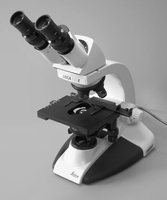
Light Microscope Structure and Function
The light microscope is an essential tool in biology for observing cells and tissues. Understanding its parts and proper use is fundamental for laboratory work.
Parts and Functions:
Eyepiece/Ocular Lens: Magnifies the image, usually 10x.
Objective Lenses: Provide additional magnification (e.g., 4x, 10x, 40x, 100x).
Nosepiece: Holds and rotates the objective lenses.
Stage: Platform where the slide is placed.
Stage Clips: Hold the slide in place.
Mechanical Stage Knob: Moves the slide on the stage.
Coarse and Fine Focus Knobs: Adjust focus; coarse for general, fine for precise focusing.
Diaphragm: Regulates light intensity.
Condenser: Focuses light onto the specimen.
Light Source: Illuminates the specimen.
Arm: Supports the microscope structure.
Focusing and Care: Start with the lowest objective, use coarse then fine focus, and handle with care to avoid damage.
Total Magnification: Multiply the eyepiece magnification by the objective lens magnification. Equation:
Key Terms:
Parfocal: Microscope remains in focus when switching objectives.
Magnification: Degree to which the image is enlarged.
Resolution: Ability to distinguish two points as separate.