 Back
BIOLOGY 1406 LAB PRACTICAL II – Step-by-Step Study Guidance
Study Guide - Smart Notes
 Tailored notes based on your materials, expanded with key definitions, examples, and context.
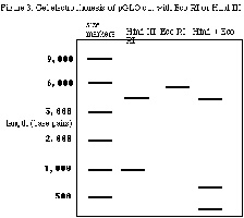
Q93. Using the figure above, what is the total length of the plasmid DNA?
Background
Topic: Gel Electrophoresis and Plasmid Mapping
This question tests your ability to interpret a gel electrophoresis image to determine the total size of a plasmid DNA molecule based on the migration of DNA fragments.
Key Terms and Concepts:
Gel Electrophoresis: A technique used to separate DNA fragments by size using an electric field.
DNA Ladder/Marker: A set of known DNA fragment sizes used as a reference.
Plasmid: A small, circular DNA molecule found in bacteria.
Restriction Digest: Cutting DNA with specific enzymes to produce fragments.
Step-by-Step Guidance
Examine the gel image and locate the lane labeled for the undigested plasmid or the lane with the largest single band (if present).
Compare the position of this band to the DNA marker (ladder) on the left side of the gel. The marker provides fragment sizes in base pairs (bp) or kilobases (kb).
Identify the band that represents the total, uncut plasmid DNA. This is usually the largest band in the undigested lane or the sum of all fragments in a complete digest.
Estimate the size of this band by matching its position to the closest band in the DNA marker lane.
Try solving on your own before revealing the answer!
Final Answer: 5,500 Kb
The largest band in the marker lane aligns with the band in the sample lane, indicating the plasmid is approximately 5,500 kilobases in length.
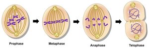
Q97. Which image represents metaphase?
Background
Topic: Mitosis Phases
This question tests your ability to identify the metaphase stage of mitosis from cell images.
Key Terms:
Metaphase: The stage of mitosis where chromosomes align at the cell's equatorial plate.
Chromosomes: Structures made of DNA and proteins, visible during cell division.
Step-by-Step Guidance
Review the images labeled 1–4. Look for the image where chromosomes are lined up in the center of the cell.
Recall that in metaphase, spindle fibers attach to the centromeres and chromosomes are most condensed and visible.
Compare each image to the description of metaphase and eliminate those that do not show chromosomes aligned at the metaphase plate.
Try solving on your own before revealing the answer!
Final Answer: Image 2
Image 2 shows chromosomes aligned at the cell's equator, characteristic of metaphase.
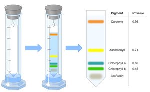
Q111. The most polar pigment is:
Background
Topic: Photosynthetic Pigments and Chromatography
This question tests your understanding of pigment polarity and how it affects migration in chromatography experiments.
Key Terms:
Polarity: The distribution of electrical charge over the atoms in a molecule.
Chromatography: A technique for separating mixtures based on differences in polarity and size.
Pigments: Molecules that absorb light, such as chlorophylls and carotenoids.
Step-by-Step Guidance
Recall that in paper chromatography, more polar pigments travel less far up the paper because they interact more with the polar paper.
Identify the pigments listed: Chlorophyll a, Chlorophyll b, xanthophyll, and carotene.
Use the Rf values (distance traveled/solvent front) to determine which pigment is most polar (lowest Rf value).
Match the pigment with the lowest Rf value to the answer choices.
Try solving on your own before revealing the answer!
Final Answer: Chlorophyll b
Chlorophyll b has the lowest Rf value, indicating it is the most polar pigment among the options.
