 Back
Biology Cell Structure, Biomolecules, and Transport Study Guide
Study Guide - Smart Notes
 Tailored notes based on your materials, expanded with key definitions, examples, and context.
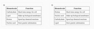
Q1. Fill in this chart with functions and examples for each Biomolecule
Background
Topic: Biomolecules and Their Functions
This question tests your understanding of the four main types of biomolecules (carbohydrates, lipids, proteins, nucleic acids) and their roles in cellular structure and function.
Key Terms:
Carbohydrates: Provide energy and structural support.
Lipids: Make up cell membranes and store energy.
Proteins: Speed up chemical reactions (enzymes), provide structure, and transport materials.
Nucleic acids: Store and transmit genetic information.
Step-by-Step Guidance
Identify the four main biomolecules: carbohydrates, lipids, proteins, and nucleic acids.
Recall the primary function of each biomolecule in cells (e.g., energy, structure, information storage).
Think of examples for each biomolecule (e.g., glucose for carbohydrates, DNA for nucleic acids).
Match each biomolecule to its function and example in the chart.
Try solving on your own before revealing the answer!
Final Answer:
Carbohydrate: Short-term energy (e.g., glucose). Lipid: Make up biological membranes (e.g., phospholipids). Protein: Speed up chemical reactions (e.g., enzymes). Nucleic acid: Store genetic information (e.g., DNA).
Each biomolecule has a unique function that is essential for cell survival and activity.
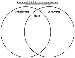
Q2. Drag and drop the features of the cell, to the correct part of the venn diagram. CELL TYPES
Background
Topic: Prokaryotic vs. Eukaryotic Cells
This question tests your ability to distinguish between prokaryotic and eukaryotic cell features.
Key Terms:
Prokaryotic: No nucleus, simple structure, includes bacteria.
Eukaryotic: Has a nucleus, complex structure, includes plants, animals, fungi, and protists.
Both: Plasma membrane, cytoplasm, ribosomes, DNA.
Step-by-Step Guidance
List cell features provided (e.g., nucleus, plasma membrane, DNA, cytoplasm, ribosomes, mitochondria, plasmid, pili, cell wall, nucleoid).
Determine which features are unique to prokaryotes (e.g., plasmid, pili, nucleoid).
Identify features unique to eukaryotes (e.g., nucleus, mitochondria).
Place features found in both cell types in the center of the Venn diagram (e.g., plasma membrane, cytoplasm, ribosomes, DNA).
Try solving on your own before revealing the answer!
Final Answer:
Prokaryotic: Plasmid, pili, nucleoid, cell wall (in most). Eukaryotic: Nucleus, mitochondria. Both: Plasma membrane, cytoplasm, ribosomes, DNA.
Understanding these features helps you distinguish cell types and their evolutionary relationships.
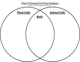
Q3. Drag and drop the features of the cell, to the correct part of the venn diagram. Differences between the plant and animal cell
Background
Topic: Plant vs. Animal Cell Structure
This question tests your knowledge of the similarities and differences between plant and animal cells.
Key Terms:
Plant cells: Cell wall, chloroplast, large central vacuole.
Animal cells: Centrioles, lysosomes (sometimes).
Both: Nucleus, mitochondria, cytoplasm, plasma membrane, ribosomes.
Step-by-Step Guidance
List cell features (e.g., cytoplasm, DNA, ribosomes, plasma membrane, cell wall, vacuole, chloroplast, nucleus, mitochondria, centrioles).
Identify features unique to plant cells (e.g., cell wall, chloroplast, large vacuole).
Identify features unique to animal cells (e.g., centrioles).
Place features found in both cell types in the center of the Venn diagram (e.g., nucleus, mitochondria, cytoplasm, plasma membrane, ribosomes).
Try solving on your own before revealing the answer!
Final Answer:
Plant: Cell wall, chloroplast, large vacuole. Animal: Centrioles. Both: Nucleus, mitochondria, cytoplasm, plasma membrane, ribosomes, DNA.
Recognizing these features helps you understand cell specialization and function.
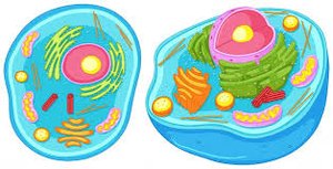
Q4. Eukaryote or Prokaryote? Animal Cell or Plant Cell
Background
Topic: Cell Classification
This question tests your ability to identify cell types based on structural features.
Key Terms:
Eukaryote: Has a nucleus.
Prokaryote: No nucleus.
Animal cell: No cell wall, no chloroplast.
Plant cell: Cell wall, chloroplast.
Step-by-Step Guidance
Look for the presence of a nucleus to determine if the cell is eukaryotic.
Check for cell wall and chloroplasts to distinguish between plant and animal cells.
Use color or structural clues (e.g., green color indicates chloroplasts, unique to plant cells).
Try solving on your own before revealing the answer!
Final Answer:
Animal cell: Has a nucleus, no cell wall or chloroplast. Plant cell: Has a nucleus, cell wall, and chloroplast.
These features are key for identifying cell types in biology.
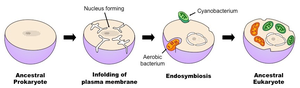
Q5. Endosymbiotic Theory: Which model on the left presents an image of how cells became complex over time?
Background
Topic: Endosymbiotic Theory
This question tests your understanding of how eukaryotic cells evolved from prokaryotic cells through endosymbiosis.
Key Terms:
Endosymbiosis: One cell engulfs another, leading to complex cells.
Eukaryotic cells: Contain organelles like mitochondria and chloroplasts, believed to have originated from prokaryotes.
Step-by-Step Guidance
Examine the models for evidence of cell complexity (e.g., organelles, nucleus).
Identify the sequence showing a prokaryote engulfing another cell.
Relate the model to the scientific theory explaining eukaryotic cell evolution.
Try solving on your own before revealing the answer!
Final Answer:
Model B shows the process of endosymbiosis, where ancestral prokaryotes become complex eukaryotes.
This supports the endosymbiotic theory, explaining the origin of mitochondria and chloroplasts.
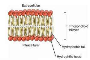
Q6. What structure does a cell have, that gives it the function of controlling materials that enter and exit the cell to maintain homeostasis? And what biomolecule helps with this function?
Background
Topic: Cell Membrane and Biomolecules
This question tests your understanding of how the cell membrane regulates transport and which biomolecules are involved.
Key Terms and Formula:
Cell membrane (plasma membrane): Controls entry and exit of substances.
Phospholipid: Main biomolecule forming the membrane.
Homeostasis: Maintaining stable internal conditions.
Step-by-Step Guidance
Identify the cell structure responsible for regulating transport (cell membrane).
Recall the biomolecule that makes up the membrane (phospholipid).
Understand how the phospholipid bilayer allows selective transport to maintain homeostasis.
Try solving on your own before revealing the answer!
Final Answer:
The cell membrane (plasma membrane) controls material movement, and phospholipids are the biomolecule responsible for this function.
This structure is essential for maintaining homeostasis in cells.
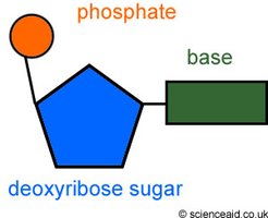
Q7. What part of the nucleotide controls cell activities by coding for proteins? What biomolecule is the structure a part of?
Background
Topic: Nucleotides and Genetic Information
This question tests your understanding of nucleotide structure and its role in protein coding.
Key Terms:
Nucleotide: Building block of nucleic acids (DNA/RNA).
Base: Part of the nucleotide that codes for proteins.
Nucleic acid: Biomolecule made of nucleotides.
Step-by-Step Guidance
Identify the three parts of a nucleotide: phosphate, sugar, base.
Recall which part codes for proteins (the base).
Determine which biomolecule the nucleotide is part of (nucleic acid).
Try solving on your own before revealing the answer!
Final Answer:
The base of the nucleotide codes for proteins, and the nucleotide is part of the nucleic acid biomolecule.
This is fundamental for genetic information and protein synthesis.
Q8. Label the parts of the viral structures on the right
Background
Topic: Viral Structure
This question tests your ability to identify and label the main parts of a virus.
Key Terms:
Nucleic acid: Genetic material (DNA or RNA).
Capsid: Protein coat surrounding the nucleic acid.
Step-by-Step Guidance
Identify the nucleic acid inside the virus.
Locate the capsid, which is the protein shell.
Label each part accordingly.
Try solving on your own before revealing the answer!
Final Answer:
Nucleic acid is the genetic material inside, and the capsid is the protein coat outside.
Viruses differ from cells in structure and function.
Q9. What is Passive Transport?
Background
Topic: Cellular Transport
This question tests your understanding of how molecules move across cell membranes without energy input.
Key Terms:
Passive transport: Movement from high to low concentration, no energy required.
Types: Diffusion, facilitated diffusion, osmosis.
Step-by-Step Guidance
Define passive transport and its characteristics.
List the types of passive transport.
Explain how concentration gradients drive passive transport.
Try solving on your own before revealing the answer!
Final Answer:
Passive transport moves molecules from high to low concentration without energy. Types include diffusion, facilitated diffusion, and osmosis.
This process is essential for maintaining cell balance.
Q10. Circle the answer that matches the following definition and use the model here: the cell transports molecules out of the cell by expelling them in an energy using process.
Background
Topic: Active Transport
This question tests your understanding of how cells use energy to move molecules out, specifically through exocytosis.
Key Terms:
Active transport: Requires energy (ATP).
Exocytosis: Expelling molecules from the cell using vesicles.
Step-by-Step Guidance
Identify the process described (energy required, molecules expelled).
Recall the term for this process (exocytosis).
Understand how vesicles and the plasma membrane are involved.
Try solving on your own before revealing the answer!
Final Answer:
Exocytosis is the process where cells use energy to expel molecules.
This is a form of active transport.
Q11. Circle the answer that matches the following definition: only refers to the movement of water.
Background
Topic: Osmosis
This question tests your understanding of osmosis, which is the movement of water across a membrane.
Key Terms:
Osmosis: Movement of water from high to low concentration across a membrane.
Step-by-Step Guidance
Recall the definition of osmosis.
Identify that osmosis is specific to water movement.
Distinguish osmosis from other types of transport.
Try solving on your own before revealing the answer!
Final Answer:
Osmosis refers only to the movement of water across a membrane.
This is a key concept in cell biology.
Q12. What component of the cell membrane allows for the transport seen in the model on the left and what biomolecule is it?
Background
Topic: Cell Membrane Structure and Transport
This question tests your understanding of how the phospholipid bilayer enables transport in and out of the cell.
Key Terms:
Phospholipid bilayer: Main component of cell membrane.
Transport: Movement of molecules via vesicles (endocytosis/exocytosis).
Step-by-Step Guidance
Identify the cell membrane component responsible for transport (phospholipid bilayer).
Recall the biomolecule involved (phospholipid).
Understand how the bilayer allows vesicle formation for bulk transport.
Try solving on your own before revealing the answer!
Final Answer:
The phospholipid bilayer enables transport, and phospholipids are the biomolecule involved.
This structure is crucial for cell function and communication.
