 Back
BackBiology Study Guide: Skeletons, Movement, Digestion, Nervous System, Heart, Evolution, and Reproduction
Study Guide - Smart Notes
 Tailored notes based on your materials, expanded with key definitions, examples, and context.
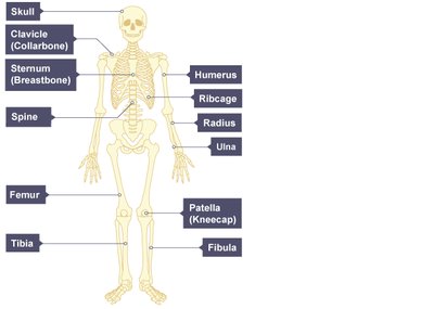
Q1. Correctly name and locate 10 different bones on the human endoskeleton.
Background
Topic: Human Skeletal System
This question tests your knowledge of the anatomy of the human skeleton, specifically the identification and location of major bones.
Key Terms:
Endoskeleton: Internal skeleton found in vertebrates.
Bone names: Skull, clavicle, sternum, spine, humerus, ribcage, radius, ulna, femur, patella, tibia, fibula.
Step-by-Step Guidance
Review the diagram of the human skeleton and note the labeled bones.
Identify the location of each bone (e.g., skull in the head, femur in the thigh).
Write down the names of 10 different bones and match them to their locations.
Check your list against the diagram to ensure accuracy.
Try solving on your own before revealing the answer!
Final Answer:
Skull, clavicle, sternum, spine, humerus, ribcage, radius, ulna, femur, patella, tibia, fibula are all correctly named and located on the diagram.
Each bone is labeled in the provided diagram, helping you visualize their positions in the human body.
Q2. State the THREE functions of the skeleton.
Background
Topic: Functions of the Skeletal System
This question tests your understanding of the main roles the skeleton plays in the human body.
Key Terms:
Muscle attachment
Protection
Support
Step-by-Step Guidance
Recall the basic functions of the skeleton: providing structure, protecting organs, and serving as attachment points for muscles.
Think about examples for each function (e.g., ribs protect the heart and lungs).
Write down the three main functions.
Try solving on your own before revealing the answer!
Final Answer:
Muscle attachment, protection, and support are the three primary functions of the skeleton.
These functions are essential for movement, safety, and maintaining body shape.
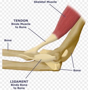
Q3. Describe the role a tendon plays at a joint.
Background
Topic: Musculoskeletal System
This question tests your understanding of how muscles and bones interact at joints.
Key Terms:
Tendon: Connects muscle to bone.
Joint: Where two bones meet.
Step-by-Step Guidance
Recall the definition of a tendon and its function.
Think about how tendons transmit force from muscle contraction to bone movement.
Describe the connection and its importance for movement.
Try solving on your own before revealing the answer!
Final Answer:
A tendon attaches a muscle to a bone, allowing muscle contractions to move bones at a joint.
This is essential for movement and stability.
Q4. Describe the role a ligament plays at a joint.
Background
Topic: Musculoskeletal System
This question tests your understanding of joint stability and the function of ligaments.
Key Terms:
Ligament: Connects bone to bone.
Joint: Where two bones meet.
Step-by-Step Guidance
Recall the definition of a ligament and its function.
Think about how ligaments stabilize joints by holding bones together.
Describe the connection and its importance for joint stability.
Try solving on your own before revealing the answer!
Final Answer:
A ligament attaches bone to bone at a joint, stabilizing the joint and preventing excessive movement.
This is crucial for maintaining proper joint function and preventing injury.
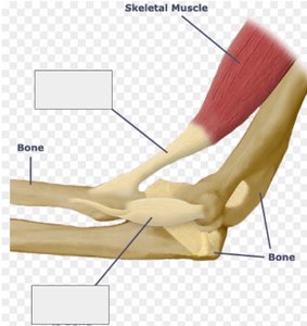
Q5. Can you label a tendon and a ligament in the diagram below?
Background
Topic: Musculoskeletal System Anatomy
This question tests your ability to visually identify and label tendons and ligaments in a joint diagram.
Key Terms:
Tendon: Muscle to bone connection.
Ligament: Bone to bone connection.
Step-by-Step Guidance
Examine the diagram and locate the structures connecting muscle to bone (tendon) and bone to bone (ligament).
Label the tendon and ligament based on their positions and connections.
Check your labels against the definitions provided.
Try solving on your own before revealing the answer!
Final Answer:
The tendon is labeled as the structure connecting muscle to bone, and the ligament is labeled as the structure connecting bone to bone.
Refer to the diagram for visual confirmation.
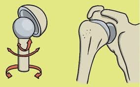
Q6. Correctly identify examples of ball and socket synovial joints.
Background
Topic: Types of Synovial Joints
This question tests your knowledge of joint types and their locations in the body.
Key Terms:
Ball and socket joint: Allows movement in multiple directions.
Synovial joint: Freely movable joint with a synovial cavity.
Step-by-Step Guidance
Recall the definition and characteristics of ball and socket joints.
Identify examples in the human body (e.g., hip, shoulder).
Write down the examples and describe their movement capabilities.
Try solving on your own before revealing the answer!
Final Answer:
Hip and shoulder joints are examples of ball and socket synovial joints.
These joints allow for a wide range of movement.
Q7. Correctly identify examples of hinge synovial joints.
Background
Topic: Types of Synovial Joints
This question tests your knowledge of hinge joints and their locations in the body.
Key Terms:
Hinge joint: Allows movement in one direction (flexion and extension).
Synovial joint: Freely movable joint with a synovial cavity.
Step-by-Step Guidance
Recall the definition and characteristics of hinge joints.
Identify examples in the human body (e.g., knee, elbow).
Write down the examples and describe their movement capabilities.
Try solving on your own before revealing the answer!
Final Answer:
Knee and elbow joints are examples of hinge synovial joints.
These joints allow for bending and straightening movements.
Q8. Describe how muscles work in pairs in the arm to allow movement using the terms contract and relax.
Background
Topic: Muscular System and Movement
This question tests your understanding of antagonistic muscle pairs and their role in movement.
Key Terms:
Contract: Muscle shortens and pulls on bone.
Relax: Muscle lengthens and allows movement.
Antagonistic pairs: Muscles that work together to produce movement.
Step-by-Step Guidance
Recall the concept of antagonistic muscle pairs (e.g., biceps and triceps).
Describe what happens when one muscle contracts and the other relaxes.
Explain how this allows movement at the joint (e.g., lifting the forearm).
Think about the importance of coordination between muscle pairs.
Try solving on your own before revealing the answer!
Final Answer:
When the biceps contract, the triceps relax, allowing the forearm to lift. When the triceps contract, the biceps relax, allowing the arm to straighten.
This coordination enables smooth and controlled movement.
