 Back
BackLESSON 6 Blood and Circulation: Structure, Function, and Regulation
Study Guide - Smart Notes
 Tailored notes based on your materials, expanded with key definitions, examples, and context.
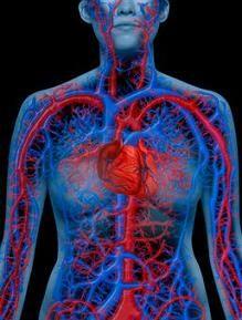
Blood and Circulation
Introduction to Circulatory Systems
The circulatory system is essential for transporting nutrients, gases, hormones, and waste products throughout the body. It consists of three main components: a circulatory fluid, a set of interconnecting vessels, and a muscular pump (the heart). There are different types of circulatory systems found in animals, including open and closed systems.
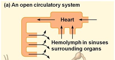
Open circulatory system: Circulatory fluid (hemolymph) bathes organs directly and is not always contained within vessels.
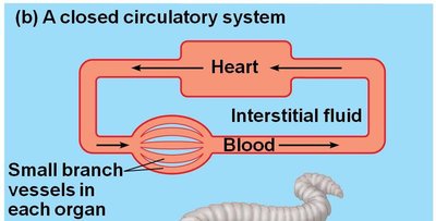
Closed circulatory system: Blood is confined to vessels and is distinct from the interstitial fluid. This system can be further divided into single and double circulation.
Types of Circulatory Systems
Open System: Found in arthropods and most mollusks. Hemolymph is pumped by the heart into body cavities, where it bathes organs directly.
Closed System: Found in annelids, cephalopods, and all vertebrates. Blood circulates within vessels, allowing for higher pressure and more efficient transport.
Single vs. Double Circulation
Single circulation: Blood passes through the heart once in each complete circuit (e.g., fish).
Double circulation: Blood passes through the heart twice in each circuit—once for pulmonary (lung) circulation and once for systemic (body) circulation (e.g., mammals, birds).
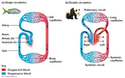
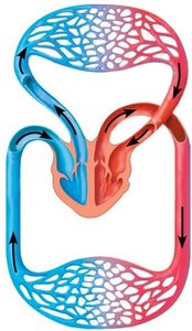
Advantage of double circulation: Oxygen-rich blood is delivered to organs at high pressure and speed, supporting higher metabolic rates.
The Heart: Structure and Function
Anatomy of the Heart
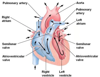
The heart is a muscular organ divided into four chambers: two atria and two ventricles. The right side pumps blood to the lungs (pulmonary circuit), while the left side pumps blood to the rest of the body (systemic circuit).
Atria: Thin-walled chambers that receive blood and pump it into the ventricles.
Ventricles: Thick-walled chambers that pump blood to the lungs or the body. The left ventricle has more muscle tissue to generate higher pressure.
Valves: Atrioventricular and semilunar valves prevent backflow of blood.
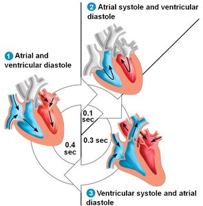
The Cardiac Cycle
The cardiac cycle consists of alternating periods of contraction (systole) and relaxation (diastole). One complete cycle takes about 0.8 seconds (75 beats per minute).
Systole: Contraction phase; blood is pumped out of the chambers.
Diastole: Relaxation phase; chambers fill with blood.
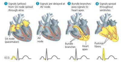
Control of Heart Rhythm
The heart's rhythmic contractions are coordinated by specialized cardiac muscle cells:
Sinoatrial (SA) node: Acts as the pacemaker, initiating the heartbeat and causing atrial contraction.
Atrioventricular (AV) node: Delays the signal, allowing the ventricles to fill before contracting.
Bundle of His and Purkinje fibers: Rapidly conduct the signal to the ventricles, ensuring coordinated contraction.
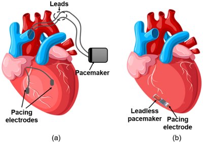
Heart Arrhythmias and Pacemakers
Arrhythmias are disorders of heart rhythm, such as tachycardia (abnormally fast heart rate). Treatments include medication (e.g., beta-blockers) and ablation procedures. Artificial pacemakers can be used to regulate heart rhythm.
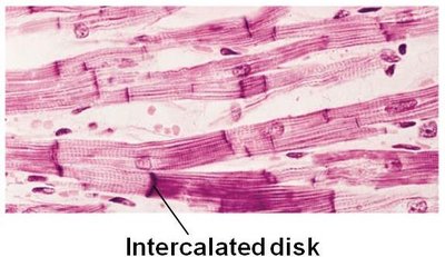
Cardiac Muscle Structure
Cardiac muscle cells are striated and connected by intercalated discs, which contain gap junctions for rapid communication. This allows the heart to contract as a functional syncytium (single unit).
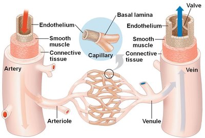
Blood Vessels: Structure and Function
Types of Blood Vessels
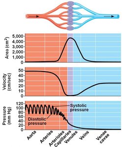
Arteries: Carry blood away from the heart to organs; have thick, muscular walls to withstand high pressure.
Capillaries: Microscopic vessels where exchange of substances occurs between blood and tissues; walls are one cell thick.
Veins: Return blood from organs to the heart; have thinner walls and valves to prevent backflow.
Blood Flow Velocity and Pressure
Blood flow is fastest in arteries and slowest in capillaries, allowing time for exchange of materials. Blood pressure decreases as blood moves through the circulatory system.
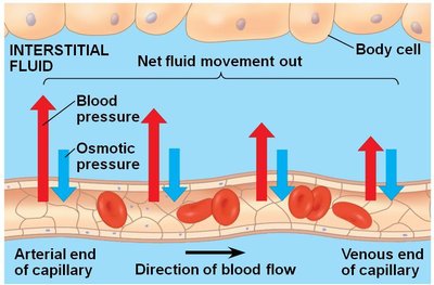
Capillary Function and Fluid Exchange
Capillaries facilitate the exchange of gases, nutrients, and waste products between blood and tissues. Blood pressure and osmotic pressure determine the direction of fluid movement.
Blood pressure: Drives fluid out of capillaries at the arterial end.
Osmotic pressure: Draws fluid back into capillaries at the venous end.
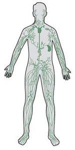
The Lymphatic System
The lymphatic system returns excess fluid and proteins from tissues to the blood. Lymph vessels are similar to veins and contain lymph nodes, which filter lymph and play a role in immune defense.
Blood: Composition and Function
Components of Blood
Plasma (55%): Mostly water (90%), ions, plasma proteins (albumin, antibodies, fibrinogen), nutrients, waste products, gases, and hormones.
Cellular elements (45%): Leukocytes (white blood cells), erythrocytes (red blood cells), and thrombocytes (platelets).
Plasma vs. Serum
Plasma: Contains clotting factors such as fibrinogen.
Serum: The fluid remaining after blood has clotted and clotting factors have been removed.
Blood Clotting
Blood clotting involves a cascade of enzymatic reactions that convert fibrinogen to fibrin, forming a clot. Platelets, damaged cells, and plasma factors (including calcium and vitamin K) are involved in this process.
Platelet plug formation
Fibrin clot formation
Key equation:
Blood Cells
Leukocytes (White Blood Cells): Variable in form; function in immune defense.
Thrombocytes (Platelets): Small cell fragments; function in blood clotting.
Erythrocytes (Red Blood Cells): Biconcave shape increases surface area; lack a nucleus to maximize hemoglobin content for oxygen transport.
Hemoglobin: The protein in erythrocytes responsible for oxygen transport.
Summary Table: Circulatory System Components
Component | Structure | Function |
|---|---|---|
Heart | Four chambers (2 atria, 2 ventricles) | Pumps blood through pulmonary and systemic circuits |
Arteries | Thick, muscular walls | Carry blood away from the heart |
Capillaries | One cell thick | Exchange of substances with tissues |
Veins | Thinner walls, valves | Return blood to the heart |
Blood | Plasma, erythrocytes, leukocytes, thrombocytes | Transport, immune defense, clotting |
Lymphatic system | Lymph vessels, lymph nodes | Returns fluid to blood, immune function |
Additional info:
The cardiac cycle and electrical conduction system are critical for maintaining coordinated heart contractions and efficient blood flow.
Blood pressure is regulated by the diameter of blood vessels and the force of heart contractions.
Disorders of the circulatory system include hypertension, atherosclerosis, and arrhythmias.
