 Back
BackBreathing and Ventilation Mechanisms in Vertebrates
Study Guide - Smart Notes
 Tailored notes based on your materials, expanded with key definitions, examples, and context.
Breathing and Ventilation in Vertebrates
Overview of Breathing Mechanisms
Breathing, or pulmonary ventilation, is the process by which air is moved into and out of the lungs to facilitate gas exchange. Different vertebrate groups have evolved distinct mechanisms for ventilating their lungs, including positive pressure breathing in amphibians, unidirectional airflow in birds, and negative pressure breathing in mammals.
Mechanisms of Breathing in Different Vertebrates
Breathing in Amphibians
Amphibians, such as frogs, utilize positive pressure breathing to ventilate their lungs. This method involves actively pushing air into the lungs by raising the floor of the oral cavity.
Inhalation: Muscles lower the floor of the oral cavity, drawing air in through the nostrils.
Air Movement: With nostrils and mouth closed, the floor rises, forcing air into the lungs via the trachea.
Exhalation: Air is expelled by the elastic recoil of the lungs and compression of the body wall.
Special Cases: During displays, male frogs may disrupt this cycle by inflating themselves without exhaling.
Breathing in Birds
Birds have a highly efficient respiratory system that uses unidirectional airflow and air sacs to maximize gas exchange. Air passes through the lungs in one direction, preventing the mixing of fresh and spent air.
Air Sacs: Located anterior and posterior to the lungs, these act as bellows to move air.
Parabronchi: Tiny channels in the lungs where gas exchange occurs.
Two-Cycle Process: Complete passage of a breath through the system requires two inhalations and two exhalations.
Efficiency: Fresh air does not mix with air that has already undergone gas exchange, maintaining a high partial pressure gradient for oxygen.
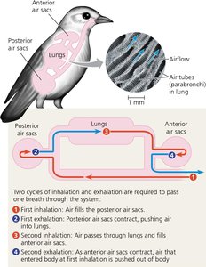
Example: Birds such as bar-headed geese can fly at high altitudes due to this efficient system, unlike mammals.
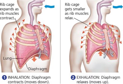
Breathing in Mammals
Mammals use negative pressure breathing, in which air is drawn into the lungs by expanding the thoracic cavity, lowering internal pressure relative to the atmosphere.
Inhalation: The diaphragm contracts and moves downward, while rib muscles contract to expand the rib cage, increasing thoracic volume and drawing air in.
Exhalation: The diaphragm and rib muscles relax, decreasing thoracic volume and pushing air out.
Alveoli: Elastic fibers in alveolar walls allow expansion and contraction with each breath. Loss of elasticity impairs gas exchange.
Residual Volume: Some air remains in the lungs after exhalation, causing mixing of fresh and residual air and reducing maximum alveolar oxygen partial pressure compared to birds.
Vital Capacity: The maximum volume of air that can be inhaled and exhaled, typically 3.4 L for women and 4.8 L for men.
Example: During exercise, additional muscles increase thoracic volume, and in some mammals, locomotion assists ventilation.
Control of Breathing in Humans
Neural Regulation of Breathing
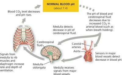
Breathing is primarily regulated by involuntary mechanisms that coordinate gas exchange with metabolic demand and blood circulation. The medulla oblongata in the brainstem contains the main control centers for breathing rhythm.
CO2 and pH Sensing: The medulla monitors the pH of cerebrospinal fluid, which reflects blood CO2 levels. Increased CO2 forms carbonic acid, lowering pH.
Feedback Mechanism: A drop in pH triggers increased depth and rate of breathing to expel CO2 and restore homeostasis.
Oxygen Sensing: Low blood O2 levels are detected by sensors in the aorta and carotid arteries, which can also stimulate increased breathing.
Stretch Receptors: Prevent overexpansion of the lungs by inhibiting further inhalation when lung tissue stretches.
Equation for CO2 and pH Regulation:
Example: During exercise, increased breathing rate and cardiac output ensure efficient O2 uptake and CO2 removal.
Key Terms and Concepts
Positive Pressure Breathing: Forcing air into the lungs by increasing pressure in the oral cavity (amphibians).
Negative Pressure Breathing: Drawing air into the lungs by expanding the thoracic cavity (mammals).
Unidirectional Airflow: Air passes in one direction through the lungs, maximizing efficiency (birds).
Tidal Volume: Volume of air inhaled or exhaled in a normal breath.
Vital Capacity: Maximum volume of air that can be moved in and out of the lungs.
Residual Volume: Air remaining in the lungs after maximal exhalation.
Medulla Oblongata: Brain region controlling breathing rhythm.
Summary Table: Comparison of Breathing Mechanisms
Group | Mechanism | Key Features |
|---|---|---|
Amphibians | Positive Pressure | Air forced into lungs by oral cavity muscles |
Birds | Unidirectional Airflow | Air sacs, parabronchi, no mixing of fresh and spent air |
Mammals | Negative Pressure | Diaphragm and rib muscles expand thoracic cavity; mixing of fresh and residual air |
Additional info: The notes above include expanded academic context on the mechanics of breathing, the role of the diaphragm, and the physiological feedback mechanisms regulating ventilation, as well as the chemical equation for CO2 buffering in blood.
