 Back
Carbohydrate Structure and Function: A Comprehensive Study Guide
Study Guide - Smart Notes
 Tailored notes based on your materials, expanded with key definitions, examples, and context.
Carbohydrate Structure and Function
Learning Objectives
Classify monosaccharides based on their structure (aldose vs ketose, by number of carbons, alpha vs beta)
Identify and name glycosidic linkages in disaccharides and polysaccharides
Compare and contrast types of polysaccharides and their functional implications
Describe the three main functions of carbohydrates
Explain why carbohydrates are the preferred energy source for all life
Monosaccharide Structure
Aldose vs Ketose
Monosaccharides are simple sugars classified by the position of their carbonyl group:
Aldose: Contains an aldehyde group (carbonyl at the end of the molecule).
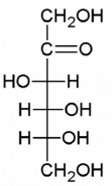
Ketose: Contains a ketone group (carbonyl within the carbon chain).
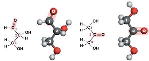
Example: Glucose is an aldose; fructose is a ketose.
Monosaccharide Isomers
Isomers are molecules with the same molecular formula but different structures. In monosaccharides, isomerism can occur due to the arrangement of hydroxyl groups around asymmetric carbons.
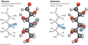
Glucose and galactose differ in the orientation of the hydroxyl group on carbon 4.
Alpha (α) vs Beta (β) Anomers
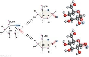
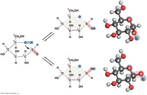
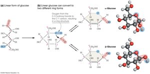
When monosaccharides form rings, the position of the hydroxyl group on the anomeric carbon (carbon 1 in glucose) determines if the sugar is alpha (α) or beta (β):
α-anomer: The OH group on the anomeric carbon is below the plane of the ring.
β-anomer: The OH group is above the plane of the ring.
Formation of the Ring Form
Monosaccharides can cyclize to form ring structures. The next-to-last hydroxyl group attacks the carbonyl carbon, forming a ring. The highest-numbered carbon remains outside the ring.
Ketoses in Ring Form
Ketose sugars, such as fructose, also form rings, but the ring closure involves different carbons compared to aldoses.
Disaccharides and Glycosidic Linkages
Glycosidic Linkages
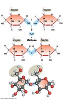
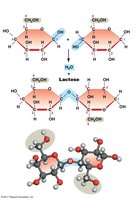
Disaccharides are formed when two monosaccharides are joined by a glycosidic linkage, which is a covalent bond formed via a dehydration reaction. The linkage is named based on the carbons involved and the anomeric configuration (α or β).
Example: α-1,4-glycosidic linkage joins the 1st carbon of one sugar (in α configuration) to the 4th carbon of another.
Polysaccharides
Starch and Glycogen
Starch (plants) and glycogen (animals) are storage polysaccharides composed of α-glucose monomers. Their structure and branching patterns affect their function:
Starch: Mixture of amylose (unbranched, α-1,4 linkages) and amylopectin (branched, α-1,6 linkages).
Glycogen: Highly branched (more α-1,6 linkages than amylopectin), allowing rapid release of glucose.
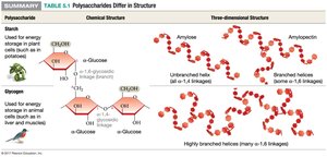
Function: Both serve as energy storage molecules, but glycogen's higher branching allows for faster mobilization in animals.
Cellulose
Cellulose is a structural polysaccharide found in plant cell walls. It consists of β-glucose monomers linked by β-1,4-glycosidic bonds, forming straight, unbranched chains that hydrogen bond to each other for strength.
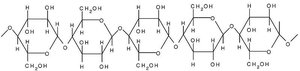
Function: Provides rigidity and structural support to plant cells.
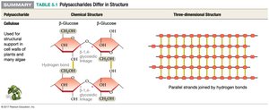
Chitin and Peptidoglycan
Chitin and peptidoglycan are structural polysaccharides similar to cellulose but with modified monosaccharides:
Chitin: Found in fungal cell walls and exoskeletons of arthropods; composed of N-acetylglucosamine (NAG) units.
Peptidoglycan: Found in bacterial cell walls; consists of alternating NAG and N-acetylmuramic acid (NAM) units, cross-linked by peptides.
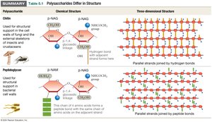
Functions of Carbohydrates
Energy Storage
Carbohydrates, especially glucose, are the primary energy source for most organisms. Glucose is metabolized via cellular respiration to produce ATP, the energy currency of the cell.
Equation:
Structural Support
Polysaccharides such as cellulose, chitin, and peptidoglycan provide structural integrity to cells and organisms by forming strong fibers or sheets stabilized by hydrogen bonds or peptide cross-links.
Cell Identity and Communication
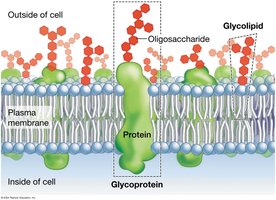
Carbohydrates attached to proteins (glycoproteins) and lipids (glycolipids) on cell surfaces serve as molecular "identification tags" for cell-cell recognition and immune response.
Carbohydrates as the Preferred Energy Source
Glucose is the preferred fuel for all life because it can be rapidly metabolized to produce ATP. Compared to fats, glucose is more readily mobilized and can be used in both aerobic and anaerobic conditions.
Advantages over fats:
Faster breakdown and ATP production
Can be used without oxygen (anaerobic glycolysis)
Essential for tissues with high energy demands (e.g., brain, muscles)
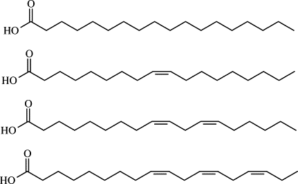
Example: During intense exercise, muscles rely on glucose for quick energy, while fats are used for long-term energy storage.
Summary Table: Major Polysaccharides
Polysaccharide | Monomer | Linkage | Function | Location |
|---|---|---|---|---|
Starch | α-glucose | α-1,4 and α-1,6 | Energy storage | Plants |
Glycogen | α-glucose | α-1,4 and α-1,6 (more branched) | Energy storage | Animals |
Cellulose | β-glucose | β-1,4 | Structural support | Plant cell walls |
Chitin | N-acetylglucosamine (NAG) | β-1,4 | Structural support | Fungi, arthropods |
Peptidoglycan | NAG and NAM | β-1,4 (with peptide cross-links) | Structural support | Bacterial cell walls |
