 Back
BackCarbon and the Molecular Diversity of Life: Biomolecules and Their Structures
Study Guide - Smart Notes
 Tailored notes based on your materials, expanded with key definitions, examples, and context.
Carbon and the Molecular Diversity of Life
Introduction to Biomolecules
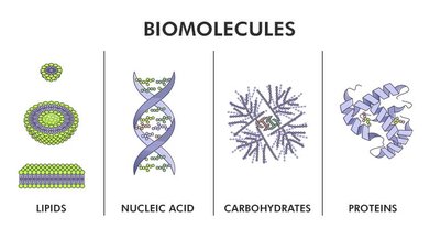
Biomolecules are the chemical compounds that form the basis of life. They are primarily organic molecules containing carbon, which allows for a vast diversity of molecular structures and functions. The four main classes of biomolecules are carbohydrates, lipids, proteins, and nucleic acids. Each class plays a unique and essential role in the structure and function of living organisms.
Carbon: The Backbone of Biological Molecules
Valence and Bonding Properties of Carbon
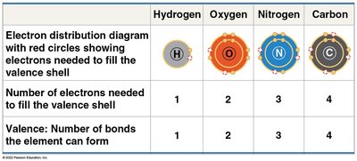
Carbon atoms have four valence electrons, allowing them to form up to four covalent bonds with other atoms. This property enables carbon to act as a versatile backbone for complex molecules, including long chains, branched structures, and rings. The diversity of carbon-based molecules underlies the complexity of biological systems.
Formation of Bonds with Carbon
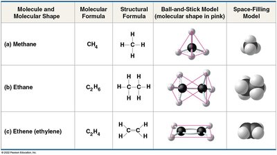
Carbon can form single, double, or triple bonds with other atoms, including hydrogen, oxygen, and nitrogen. The shape of carbon-containing molecules depends on the types of bonds formed. For example, single bonds allow for tetrahedral geometry, while double bonds create planar structures.
Carbon Skeletons and Their Variations
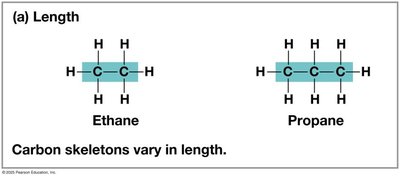
Carbon skeletons form the framework of organic molecules and can vary in length, branching, double bond position, and ring formation. These variations contribute to the diversity of organic compounds found in living organisms.
Length: Carbon chains can be short or long.
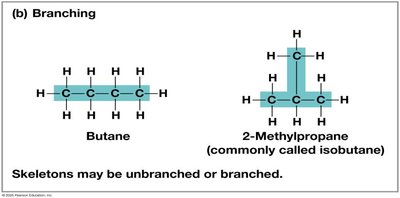
Branching: Chains may be unbranched or branched.
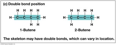
Double Bond Position: Double bonds can occur at different positions along the chain.
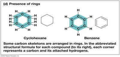
Rings: Some carbon skeletons form rings, such as cyclohexane and benzene.
Isomers: Structural Diversity
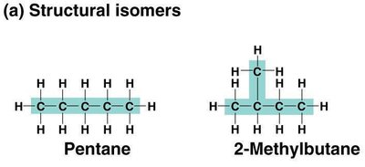
Isomers are compounds with the same molecular formula but different structures and properties. There are three main types:
Structural Isomers: Differ in the covalent arrangement of atoms.
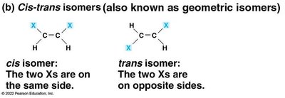
Cis-Trans (Geometric) Isomers: Differ in spatial arrangement around double bonds.
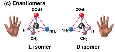
Enantiomers: Mirror images of each other, often with different biological activities.
Functional Groups
Functional groups are specific groups of atoms attached to carbon skeletons that participate in chemical reactions and confer unique properties to molecules. Examples include hydroxyl, carbonyl, carboxyl, amino, sulfhydryl, phosphate, and methyl groups. These groups are critical in determining the behavior of biomolecules.
ATP: The Energy Currency of the Cell
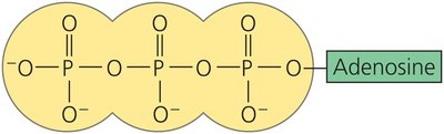
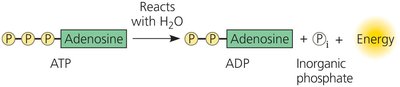
Adenosine triphosphate (ATP) is an organic molecule that stores and transfers energy within cells. ATP consists of adenosine attached to three phosphate groups. Hydrolysis of ATP releases energy used for cellular processes.
Macromolecules: Polymers and Monomers
Polymer Formation and Breakdown
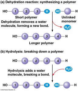
Most biomolecules are polymers, long chains made from repeating units called monomers. Polymers are synthesized by dehydration reactions (removal of water to form a bond) and broken down by hydrolysis reactions (addition of water to break a bond). Enzymes catalyze these reactions.
Carbohydrates
Monosaccharides
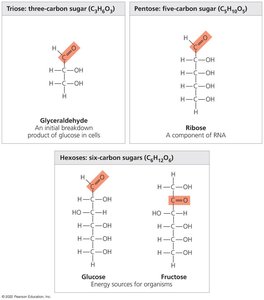
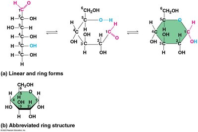
Carbohydrates are sugars and their polymers. The simplest carbohydrates are monosaccharides, such as glucose (C6H12O6). Monosaccharides are classified by the number of carbons and the position of the carbonyl group. In aqueous solutions, most monosaccharides form ring structures.
Disaccharides and Glycosidic Linkages
Disaccharides are formed when two monosaccharides are joined by a glycosidic linkage through a dehydration reaction. Sucrose (table sugar) is a common disaccharide composed of glucose and fructose.
Polysaccharides: Storage and Structural Roles
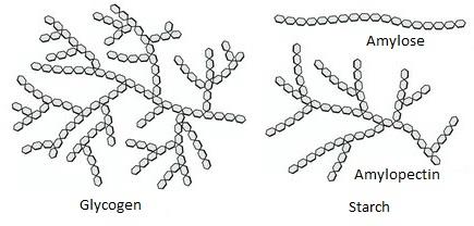
Polysaccharides are large carbohydrate polymers with storage or structural functions. Examples include:
Starch: Storage polysaccharide in plants, composed of glucose monomers.
Glycogen: Storage polysaccharide in animals, mainly in liver and muscle cells.
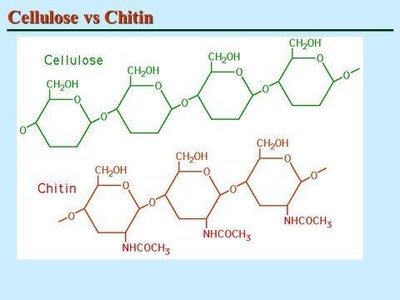
Cellulose: Structural polysaccharide in plant cell walls; most abundant organic compound on Earth.
Chitin: Structural polysaccharide in arthropod exoskeletons and fungal cell walls.
Lipids
Structure and Function of Lipids
Lipids are hydrophobic molecules that do not form true polymers. They are mainly composed of hydrocarbons and include fats, phospholipids, and steroids. Lipids serve as energy storage, structural components of cell membranes, and signaling molecules.
Fats and Fatty Acids
Fats are constructed from glycerol and three fatty acids via dehydration reactions, forming triacylglycerol (triglyceride). Fatty acids can be saturated (no double bonds, solid at room temperature) or unsaturated (one or more double bonds, liquid at room temperature).
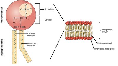
Phospholipids
Phospholipids consist of two fatty acids, a phosphate group, and glycerol. They are essential for cell membrane structure, forming a bilayer with hydrophilic heads and hydrophobic tails.
Steroids
Steroids are lipids with a carbon skeleton of four fused rings. Cholesterol is a key steroid in animal cell membranes and a precursor for other steroids such as testosterone and estrogen.
Proteins
Structure and Function of Proteins
Proteins are polymers of amino acids and account for more than 50% of the dry mass of most cells. They perform a wide range of functions, including catalysis, defense, transport, support, movement, and regulation. Each protein has a unique three-dimensional structure that determines its function.
Amino Acids and Peptide Bonds
Amino acids are organic molecules with amino and carboxyl groups, differing in their side chains (R groups). Amino acids are linked by peptide bonds to form polypeptides. The sequence of amino acids determines the protein's structure and function.
Levels of Protein Structure
Primary Structure: Linear sequence of amino acids.
Secondary Structure: Coils and folds (α-helix, β-pleated sheet) stabilized by hydrogen bonds.
Tertiary Structure: Overall 3D shape formed by interactions among R groups (hydrophobic interactions, disulfide bridges).
Quaternary Structure: Association of two or more polypeptides (e.g., hemoglobin).
Protein Structure and Disease
A single amino acid substitution can drastically affect protein function, as seen in sickle-cell disease, where a mutation in hemoglobin leads to abnormal red blood cell shape and function.
Nucleic Acids
DNA and RNA: Structure and Function
Nucleic acids store, transmit, and help express hereditary information. The two main types are deoxyribonucleic acid (DNA) and ribonucleic acid (RNA). DNA contains the genetic blueprint, while RNA is involved in protein synthesis and gene regulation.
Nucleotide Structure
Nucleotides are the monomers of nucleic acids, each consisting of a nitrogenous base, a pentose sugar, and one or more phosphate groups. DNA uses deoxyribose, while RNA uses ribose. Nitrogenous bases are classified as purines (adenine, guanine) or pyrimidines (cytosine, thymine, uracil).
DNA and RNA Polymers
Nucleotides are joined by phosphodiester linkages to form polynucleotide chains. DNA is typically double-stranded, forming a double helix with complementary base pairing (A-T, G-C). RNA is usually single-stranded but can form complex structures through internal base pairing.
Genomics and Proteomics
Genomics is the study of whole sets of genes and their interactions, while proteomics focuses on the study of entire sets of proteins. Advances in bioinformatics have accelerated research in these fields, allowing for rapid sequencing and analysis of genomes and proteomes.
DNA and Proteins as Evolutionary Tape Measures
The linear sequences of nucleotides in DNA are inherited and can be compared across individuals and species to assess evolutionary relationships. Molecular biology provides tools for understanding evolutionary kinship and genetic diversity.
