 Back
BackCarbon and the Molecular Diversity of Life
Study Guide - Smart Notes
 Tailored notes based on your materials, expanded with key definitions, examples, and context.
Chapter 3: Carbon and the Molecular Diversity of Life
Introduction to Biomolecules
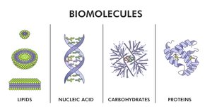
Living organisms are composed of a diverse array of organic molecules, all of which are based on the element carbon. These molecules are large, complex, and varied, forming the foundation of biological structure and function. The four main classes of biomolecules are carbohydrates, lipids, proteins, and nucleic acids.
3.1 Carbon Atoms Can Form Diverse Molecules by Bonding to Four Other Atoms
The Unique Properties of Carbon
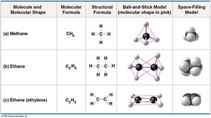
Carbon is unparalleled in its ability to form large, complex, and diverse molecules. This versatility arises from its four valence electrons, allowing it to form four covalent bonds with a variety of atoms, including other carbon atoms. This property enables the construction of an immense variety of molecular architectures essential for life.
Valence: The number of covalent bonds an atom can form, determined by the number of unpaired electrons in its outer shell.
Tetrahedral Geometry: When carbon forms four single bonds, the molecule adopts a tetrahedral shape. Double bonds create planar (flat) regions in molecules.
Formation of Bonds with Carbon
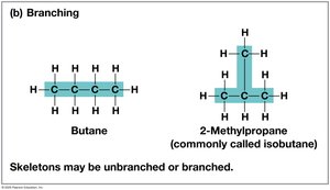
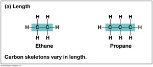
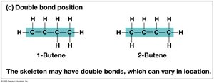
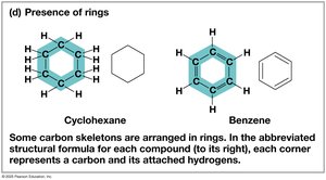
Carbon commonly bonds with hydrogen, oxygen, and nitrogen, as well as with other carbon atoms, forming the backbone of organic molecules. These backbones can be straight, branched, or arranged in rings, and may include single, double, or triple bonds.
Hydrocarbons: Molecules consisting only of carbon and hydrogen. They are nonpolar and hydrophobic, and serve as energy sources in fats.
Carbon Skeletons: The chain of carbon atoms in an organic molecule, which can vary in length, branching, double bond position, and ring formation.
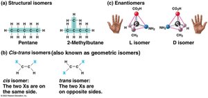
Isomers: Structural Diversity in Organic Molecules
Isomers are compounds with the same molecular formula but different structures and properties. There are three main types:
Structural Isomers: Differ in the covalent arrangement of atoms.
Cis-trans (Geometric) Isomers: Differ in spatial arrangement around double bonds; cis isomers have groups on the same side, trans on opposite sides.
Enantiomers: Mirror images of each other, often with only one form biologically active.
Functional Groups
Functional groups are specific groups of atoms attached to carbon skeletons that participate in chemical reactions and confer specific properties to molecules. Seven functional groups are especially important in biological chemistry: hydroxyl, carbonyl, carboxyl, amino, sulfhydryl, phosphate, and methyl.
Functional groups are not unique to one biomolecule and can be found in many different molecules.
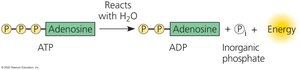
ATP: The Energy Currency of the Cell
Adenosine triphosphate (ATP) is an organic molecule consisting of adenosine attached to three phosphate groups. ATP stores potential energy in its phosphate bonds, which is released upon hydrolysis to power cellular processes.

3.2 Macromolecules Are Polymers, Built from Monomers
Polymers and Monomers
Most macromolecules are polymers, long molecules built from repeating units called monomers. The four classes of macromolecules are constructed from different types of monomers:
Carbohydrates: Monosaccharides
Proteins: Amino acids
Nucleic acids: Nucleotides
Lipids: Not true polymers, but assembled from smaller molecules
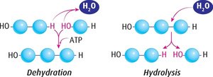
Polymers are synthesized by dehydration reactions (removal of water to form a bond) and broken down by hydrolysis reactions (addition of water to break a bond).
Carbohydrates
Monosaccharides
Monosaccharides are the simplest carbohydrates, serving as fuel and building blocks for more complex sugars. They have the general formula (CH2O)n, with glucose (C6H12O6) being the most common. Monosaccharides can form rings in aqueous solutions, which is the most stable form under physiological conditions.
Disaccharides and Polysaccharides
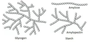
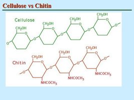
Disaccharides are formed by joining two monosaccharides via a dehydration reaction, creating a glycosidic linkage (e.g., sucrose = glucose + fructose). Polysaccharides are long chains of monosaccharides and serve storage (starch in plants, glycogen in animals) or structural (cellulose in plants, chitin in arthropods and fungi) roles.
Lipids
Structure and Function
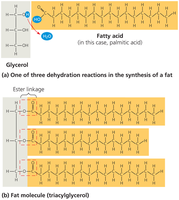
Lipids are hydrophobic molecules that do not form true polymers. Major types include fats, phospholipids, and steroids. Fats are constructed from glycerol and three fatty acids, forming triglycerides via ester linkages. Lipids are important for energy storage, membrane structure, and signaling.
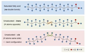
Saturated vs. Unsaturated Fatty Acids
Saturated fatty acids: No double bonds, solid at room temperature, mostly animal fats.
Unsaturated fatty acids: One or more double bonds, liquid at room temperature, mostly plant and fish fats.
Trans fats: Unsaturated fats with trans double bonds, often produced industrially.
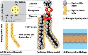
Phospholipids and Steroids
Phospholipids consist of two fatty acids, a phosphate group, and glycerol. They are amphipathic, with hydrophilic heads and hydrophobic tails, and form the basis of cell membranes as bilayers.
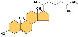
Steroids are lipids with a structure of four fused rings. Cholesterol is a key steroid, maintaining membrane fluidity and serving as a precursor for other steroids such as hormones.
Proteins
Structure and Function
Proteins are polymers of amino acids, accounting for more than 50% of the dry mass of most cells. They perform a vast array of functions, including catalysis, defense, storage, transport, communication, movement, and structural support. Each protein has a unique three-dimensional structure determined by its amino acid sequence.
Amino Acids and Polypeptides
Amino acids are organic molecules with amino and carboxyl groups, differing in their side chains (R groups). Peptide bonds link amino acids into polypeptides, which fold into functional proteins.
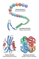
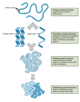
Levels of Protein Structure
Primary structure: Linear sequence of amino acids.
Secondary structure: Coils and folds (α-helix, β-pleated sheet) due to hydrogen bonding.
Tertiary structure: Overall 3D shape from interactions among R groups (hydrophobic interactions, disulfide bridges, etc.).
Quaternary structure: Association of two or more polypeptides (e.g., hemoglobin).

Protein Structure and Disease
A single amino acid substitution can drastically affect protein function, as seen in sickle-cell disease, where a change in hemoglobin leads to abnormal red blood cell shape and function.
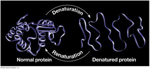
Denaturation
Protein structure can be disrupted by changes in temperature, pH, or salt concentration, leading to denaturation (loss of native structure and function). Sometimes, denaturation is reversible.
Nucleic Acids
DNA and RNA: Information Molecules
Nucleic acids store, transmit, and help express hereditary information. DNA encodes genetic instructions, while RNA is involved in protein synthesis. The flow of genetic information follows the central dogma: DNA → RNA → Protein.
Structure of Nucleic Acids
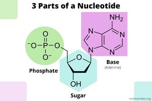
Nucleic acids are polymers (polynucleotides) made of nucleotide monomers. Each nucleotide consists of a nitrogenous base, a pentose sugar (deoxyribose in DNA, ribose in RNA), and one or more phosphate groups. Nitrogenous bases are classified as pyrimidines (C, T, U) or purines (A, G).
DNA and RNA Structure
DNA: Double helix with antiparallel strands; complementary base pairing (A-T, G-C).
RNA: Single-stranded; can form complex structures via internal base pairing; uracil replaces thymine (A-U pairing).
Genomics and Proteomics
Modern biology uses genomics (study of whole genomes) and proteomics (study of entire sets of proteins) to understand biological complexity. Bioinformatics applies computational tools to analyze large datasets, revealing evolutionary relationships and functional insights.
