 Back
Cell-Cell Interactions: Structure, Function, and Communication in Multicellular Organisms
Study Guide - Smart Notes
 Tailored notes based on your materials, expanded with key definitions, examples, and context.
Cell-Cell Interactions
Introduction to Cell-Cell Interactions
Cell-cell interactions are fundamental for the survival and function of both unicellular and multicellular organisms. In multicellular organisms, cells must communicate and cooperate to maintain homeostasis, form tissues, and respond to environmental changes. This section explores the structural and functional aspects of cell surfaces, extracellular layers, and the mechanisms by which cells connect and communicate.
The Cell Surface
Structure of the Plasma Membrane
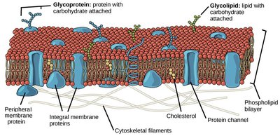
The plasma membrane is a selectively permeable barrier composed of a phospholipid bilayer embedded with proteins. These proteins can be integral (spanning the membrane) or peripheral (attached to the membrane surface). Membrane proteins regulate the transport of substances, maintain cellular environments, and anchor the cytoskeleton and extracellular structures.
Integral membrane proteins: Span the membrane and are involved in transport and signaling.
Peripheral membrane proteins: Attach to the membrane surface and play roles in signaling and maintaining cell shape.
Glycoproteins and glycolipids: Involved in cell recognition and signaling.
Protective Layers Beyond the Plasma Membrane
Most cells possess a protective layer or wall beyond the plasma membrane. This extracellular membrane (ECM) helps define cell shape, attaches cells to one another, and acts as a first line of defense against environmental stress.
Structure and Function of Extracellular Layers
Prokaryotic Cell Walls
Prokaryotes have distinct extracellular structures:
Bacteria: Cell walls are primarily composed of peptidoglycan polymers cross-linked by peptide bonds.
Archaea: Lack peptidoglycan; instead, their cell walls form a dense protein coat known as the S-layer.
Extracellular Layers in Eukaryotes
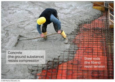
Eukaryotic cells, including plants and animals, have extracellular layers with a common organizational theme: a network of long filaments (resisting tension) embedded in a stiff ground substance (resisting compression). This structure provides both strength and flexibility.

The Primary Cell Wall in Plants
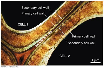
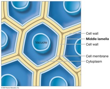
Most plant cells are surrounded by a primary cell wall, a fiber composite mainly composed of cellulose microfibrils in a matrix of gelatinous polysaccharides such as pectin. This wall defines cell shape and counteracts turgor pressure from water uptake.
Cellulose microfibrils: Provide tensile strength.
Pectin: Maintains wall moisture and flexibility.

Role of Turgor Pressure and Expansins
Turgor pressure is crucial for young, growing plant cells. Proteins called expansins disrupt cross-linking between microfibrils, allowing the cell wall to expand and the cell to grow.

The Secondary Cell Wall in Plants
Mature plant cells may secrete a secondary cell wall between the plasma membrane and the primary cell wall. Its composition varies by cell type: waxes in leaf cells and lignin in wood-forming cells, providing additional strength and protection.
The Extracellular Matrix (ECM) in Animals
Animal cells secrete a fiber composite called the extracellular matrix (ECM), which provides structural support. The ECM consists mainly of collagen fibers (resisting tension) and proteoglycans (providing a gel-like ground substance that resists compression).
Collagen: Triple-helix proteins that form strong fibrils.
Proteoglycans: Proteins with attached polysaccharides, giving cartilage its rubbery texture.
Variation and Attachment in the ECM
The amount and composition of ECM vary among tissues. For example, elastin allows lung tissue to stretch. Integrins are membrane proteins that connect the ECM to the cytoskeleton, anchoring cells and facilitating communication.

How Do Adjacent Cells Connect and Communicate?
Direct Physical Connections
Direct connections between cells are essential for maintaining tissue structure and function. These connections are especially important in epithelia, tissues that line surfaces and organs. The structures that hold cells together vary among organisms and tissue types.
Indirect Cell–Cell Attachments in Plants
In plants, the middle lamella is a gelatinous layer rich in pectins that glues adjacent cells together, forming a continuous network with the primary cell walls of neighboring cells.

Tight Junctions in Animals
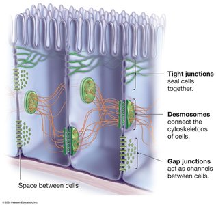
Tight junctions are cell–cell attachments composed of membrane proteins in adjacent animal cells. These proteins form chains that bind together, creating a watertight seal. Tight junctions are dynamic, can open or close in response to environmental changes, and are commonly found in epithelial tissues.
Desmosomes: Secure Adhesions
Desmosomes are strong cell–cell attachments found in animal epithelial and muscle cells. They consist of linking proteins and cytosolic anchoring proteins, with cytoskeletal intermediate filaments reinforcing the structure.
Selective Cell–Cell Adhesion
Animal cells exhibit selective adhesion, binding only to cells with compatible adhesion molecules. Experiments with sponges demonstrated that dissociated cells can reaggregate with cells of the same type, reforming functional tissues.
Discovery of Cell–Cell Adhesion Proteins
Researchers identified that different cell types have unique adhesion proteins. Cadherins are a major class of adhesion proteins in desmosomes, binding only to cadherins of the same type, ensuring specific cell–cell attachment.
Cell Communication via Cell–Cell Gaps
Gap Junctions in Animals
Gap junctions are protein channels that connect adjacent animal cells, allowing the flow of ions and small molecules. These communication portals help coordinate cellular activities and enable rapid signaling between cells.
Plasmodesmata in Plants
Plant cells are connected by plasmodesmata, membrane-lined channels that traverse the cell wall. These channels connect the plasma membranes, cytoplasm, and smooth endoplasmic reticulum of adjacent cells, dividing plant tissues into two compartments:
Symplast: Shared cytoplasm between cells.
Apoplast: The extracellular space outside the plasma membrane.
Comparison: Unicellular vs. Multicellular Organisms
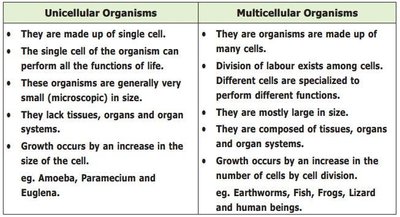
Unicellular Organisms | Multicellular Organisms |
|---|---|
Made up of a single cell | Made up of many cells |
Single cell performs all life functions | Division of labor among specialized cells |
Microscopic in size | Larger in size |
Lack tissues, organs, and organ systems | Composed of tissues, organs, and organ systems |
Growth by increase in cell size | Growth by increase in cell number via division |
Examples: Amoeba, Paramecium, Euglena | Examples: Earthworms, Fish, Frogs, Lizards, Humans |
Summary
Cell-cell interactions are essential for the structure, function, and communication of cells in multicellular organisms. The plasma membrane, extracellular layers, and specialized junctions enable cells to adhere, communicate, and coordinate activities, ensuring the integrity and adaptability of tissues and organs.
