 Back
BackCell Communication and the Cell Cycle: Key Concepts and Mechanisms
Study Guide - Smart Notes
 Tailored notes based on your materials, expanded with key definitions, examples, and context.
Cell Communication & Signaling
Cellular Messaging
Cells communicate with each other and their environment using chemical signals. These signals are often small molecules that can diffuse rapidly, enabling efficient communication. The mechanisms of cell signaling are conserved across diverse species and biological processes.
Chemical signals are the primary mode of cellular communication.
Signaling mechanisms are highly conserved and appear in many organisms.
Example: Saccharomyces cerevisiae (yeast) uses mating factors to locate and recognize opposite mating types.
External Signals and Cellular Responses
Cells convert external signals into internal responses through signal transduction pathways. These pathways involve a series of molecular interactions that ultimately lead to a specific cellular response.
Signal transduction pathways are similar in yeast and mammals.
Example: Hormone epinephrine acts on cells via a specific receptor-mediated process.
The Three Stages of Cell Signaling
Cell signaling involves three main stages, as discovered by Earl W. Sutherland:
Reception: Signal molecule binds to a receptor, causing a shape change.
Transduction: Signal is relayed through a cascade of proteins.
Response: Activation of a specific cellular process or protein.
Reception: Membrane Receptors
Reception occurs when a signaling molecule (ligand) binds to a receptor protein, often located in the plasma membrane. The binding is highly specific and induces a conformational change in the receptor.
Three main types of membrane receptors:
G protein-coupled receptors (GPCRs): Transmembrane receptors that interact with G proteins, which bind GTP. GPCRs are diverse and widespread.
Receptor tyrosine kinases (RTKs): Membrane receptors that phosphorylate tyrosines. RTKs can trigger multiple pathways and are linked to cancer when dysfunctional.
Ion channel receptors: Ligand-gated channels that open to allow ions (e.g., Na+, Ca2+) to pass through, acting as second messengers.
Intracellular Receptors
Intracellular receptors are found in the cytoplasm or nucleus. Small or hydrophobic messengers, such as steroid and thyroid hormones, can cross the membrane and activate these receptors. The activated complex often acts as a transcription factor, regulating gene expression.
Signal Transduction Pathways
Signal transduction involves a cascade of molecular interactions, often resulting in a change in protein shape. Each step amplifies and transduces the signal, leading to the final cellular response.
Protein Phosphorylation and Dephosphorylation
Phosphorylation (by kinases) and dephosphorylation (by phosphatases) regulate protein activity. This system acts as a molecular switch, controlling cellular processes.
Protein kinases: Transfer phosphate from ATP to proteins.
Protein phosphatases: Remove phosphate groups from proteins.
Phosphorylation cascades are common in signal transduction.
Second Messengers
Second messengers are small, non-protein molecules or ions that diffuse rapidly within the cell. They participate in pathways initiated by GPCRs and RTKs.
Cyclic AMP (cAMP): Produced by adenylyl cyclase from ATP in response to signals.
Calcium ions (Ca2+): Act as second messengers; their concentration is tightly regulated.
Inositol triphosphate (IP3): Also involved in calcium signaling.
Nuclear and Cytoplasmic Responses
Signal transduction pathways regulate cellular activities in the cytoplasm or nucleus. Many pathways control gene expression by activating transcription factors.
Regulation of the Response
Cellular responses to signals are regulated in several ways:
Amplification: One signal can trigger many responses.
Specificity: Different cells respond differently to the same signal.
Efficiency: Scaffolding proteins enhance response efficiency.
Termination: Inactivation mechanisms ensure signals are not perpetuated indefinitely.
Termination of the Signal
Signal termination is essential for proper cell function. When ligand concentration decreases, receptors become unbound and revert to an inactive state.
Apoptosis: Programmed Cell Death
Cells that are damaged, infected, or no longer needed undergo apoptosis, a process regulated by multiple signaling pathways.
The Cell Cycle
Binary Fission in Bacteria
Bacterial cell division occurs via binary fission, a process involving chromosome replication and segregation, resulting in two daughter cells.
Chromosome replication begins.
Each copy moves to opposite ends of the cell.
Replication finishes.
Two daughter cells are produced.
Cellular Division in Eukaryotes
Cell division is essential for reproduction, growth, and repair in multicellular organisms. It is a fundamental part of the cell cycle, which encompasses the life of a cell from formation to division.
Genome: All DNA in a cell.
Prokaryotes: Single DNA molecule.
Eukaryotes: Multiple DNA molecules packaged into chromosomes.
Chromatin: DNA-protein complex in eukaryotic chromosomes.
Somatic cells: Two sets of chromosomes.
Gametes: One set of chromosomes.
Chromosomes: Structure and Organization
Each duplicated chromosome consists of two sister chromatids joined by cohesion proteins. The centromere is the region where chromatids are most closely attached.
Phases of the Cell Cycle
The cell cycle consists of the Mitotic (M) phase and Interphase. Interphase is subdivided into G1, S, and G2 phases. Chromosome duplication occurs only during the S phase.
Mitotic (M) phase: Includes mitosis (chromosome distribution) and cytokinesis (cytoplasm division).
Interphase: Cell growth and chromosome copying.
Sub-phases:
G1 phase: Growth and metabolic activity.
S phase: DNA synthesis and growth.
G2 phase: Preparation for cell division.
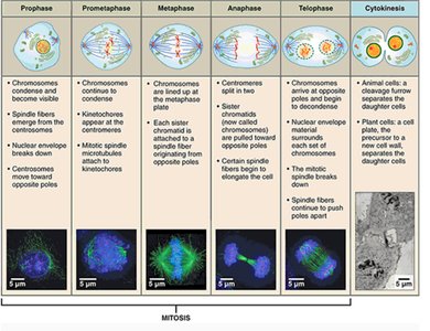
Mitosis: Stages and Key Events
Mitosis is the process by which eukaryotic cells divide their chromosomes into two daughter nuclei. It consists of several stages:
Prophase: Chromosomes condense and become visible; spindle fibers form; nuclear envelope breaks down.
Prometaphase: Chromosomes continue to condense; kinetochores form; spindle fibers attach to kinetochores.
Metaphase: Chromosomes align at the metaphase plate; spindle fibers connect to kinetochores.
Anaphase: Sister chromatids separate and move to opposite poles; microtubules shorten.
Telophase: Chromosomes arrive at poles; nuclear envelope reforms; spindle fibers disassemble.
Cytokinesis: Division of cytoplasm; cleavage furrow forms in animal cells, cell plate forms in plant cells.
The Cell Cycle Clock: Cyclins and Cyclin-Dependent Kinases
Cell cycle progression is regulated by cyclins and cyclin-dependent kinases (Cdks). Cyclins fluctuate in concentration, and Cdks are active only when bound to cyclins. MPF (maturation-promoting factor) is a cyclin-Cdk complex that triggers entry into the M phase.
Kinase: Adds phosphate to proteins.
Phosphatase: Removes phosphate from proteins.
Cell Cycle Checkpoints: Internal and External Signals
Checkpoints are control mechanisms that ensure proper cell cycle progression. Signals from within and outside the cell are registered at these checkpoints.
Three main checkpoints: G1, G2, and M phases.
G1 checkpoint is often the most important; cells that do not pass enter G0 (nondividing state).
Internal signal: Chromosomes must be properly attached to the spindle before anaphase.
External signal: Growth factors stimulate cell division; normal cells require anchorage and exhibit density-dependent inhibition.
Loss of Cell Cycle Controls in Cancer Cells
Cancer cells bypass normal cell cycle controls, leading to uncontrolled division. They may produce their own growth factors, ignore external signals, or have abnormal control systems. Tumors form when cancer cells are not eliminated by the immune system.
Benign tumors: Remain at the original site.
Malignant tumors: Invade surrounding tissues and can metastasize.
Cancer cells often lose the ability to repair DNA damage.
Key characteristics: No need for normal growth signals, loss of anchorage dependence, loss of density-dependent inhibition.
Additional info: The included image provides a visual summary of the stages of mitosis, reinforcing the textual explanation of mitotic events and cellular structures.
