 Back
BackCell Communication, Signal Transduction, Feedback Mechanisms, and Cell Cycle Regulation
Study Guide - Smart Notes
 Tailored notes based on your materials, expanded with key definitions, examples, and context.
Cell Communication
Ligands, Receptors, and Target Cells
Cell communication is essential for coordinating cellular activities and responses to environmental signals. The process involves three main components:
Ligand: A molecule that binds to a specific site on a receptor, acting as a 'key'.
Receptor: A protein, typically located on the cell surface or inside the cell, that binds to the ligand ('lock').
Target Cell: The cell possessing the receptor; upon ligand binding, it initiates a specific response.
Sequence: Ligand → Receptor → Target Cell
Methods of Cellular Communication
Direct Cell-to-Cell (Juxtacrine): Requires physical contact between cells. Examples include gap junctions (animal cells), plasmodesmata (plant cells), and cell-cell recognition (both).
Local Signaling:
Autocrine: Cell releases a ligand that binds to its own receptors.
Paracrine: Ligand affects nearby cells.
Synaptic: Specialized paracrine signaling in neurons.
Quorum Sensing: Bacteria release autoinducers to coordinate group behaviors based on population density.
Long Distance Signaling:
Endocrine: Hormones travel through the bloodstream to distant cells.
Nervous System: Neurons transmit electrical signals over long distances; neurotransmitter release at synapses is local.
Example: Endocrine signaling in humans involves hormones such as insulin regulating blood glucose levels.
Signal Transduction
Major Steps of Signal Transduction Pathway
Reception: Detection of a ligand by a receptor.
Transmembrane Receptors:
G Protein Coupled Receptor (GPCR): Ligand binding activates G protein, triggering downstream signaling.
Ligand-Gated Channel: Ligand binding opens channel, allowing ion flow.
Receptor Tyrosine Kinase (RTK): Ligand binding causes dimerization and phosphorylation, activating signaling proteins.
Intracellular Receptors: Located in the cytoplasm or nucleus; respond to hydrophobic ligands.
Transduction: Conversion and amplification of the signal inside the cell.
Kinase: Enzyme that adds phosphate groups (phosphorylation).
Phosphatase: Enzyme that removes phosphate groups (dephosphorylation).
Phosphorylation Cascade: Sequential activation of kinases, amplifying the signal.
Second Messengers: Small molecules (e.g., cAMP, Ca2+) that amplify and transmit signals.
Response: Cellular changes resulting from the signal.
Cytoplasmic Response: Rapid changes, such as glycogen breakdown.
Nuclear Response: Changes in gene expression via transcription factors.
Example Pathway: In the cAMP pathway, a hormone binds to a GPCR, activating adenylate cyclase, which converts ATP to cAMP. cAMP then activates protein kinase A, leading to cellular responses.
Feedback Mechanisms
Positive vs. Negative Feedback
Negative Feedback: Reduces the original stimulus, maintaining homeostasis (e.g., body temperature regulation).
Positive Feedback: Amplifies the original stimulus, driving processes to completion (e.g., blood clotting).
Cell Cycle
Key Terms and Structures
Chromatin: Loose DNA-protein complex in the nucleus.
Chromatid/Sister Chromatids: Chromatid is one half of a duplicated chromosome; sister chromatids are two identical copies.
Chromosome: Highly condensed DNA-protein structure.
Centromere: Region joining sister chromatids.
Centrosome: Organelle organizing microtubules and mitotic spindle.
Somatic Cell Division
Somatic cells (diploid) divide by mitosis to produce identical daughter cells.
If a somatic cell has 50 chromosomes, each daughter cell will also have 50 chromosomes.
Organization of Eukaryotic DNA
Hierarchy: DNA → nucleosome → chromatin → chromatid → chromosome (sister chromatids)
Mitotic Spindle Components
Centrosomes: Anchor and organize spindle microtubules.
Spindle Microtubules: Attach to chromosomes and pull them apart.
Asters: Star-shaped microtubule arrays, help position spindle apparatus.
Kinetochores
Protein complexes on centromeres; attach chromosomes to spindle fibers.
Critical during metaphase (alignment) and anaphase (separation).
Cell Division Phases
Interphase:
G1: Cell growth, protein synthesis.
S: DNA replication, centrosome duplication.
G2: Further growth, DNA error checking and repair.
Mitotic (M) Phase:
Mitosis: Division of genetic material into two nuclei.
Prophase: Chromatin condenses.
Prometaphase: Nuclear envelope disappears, spindle fibers attach.
Metaphase: Chromosomes align at metaphase plate.
Anaphase: Sister chromatids pulled apart.
Telophase: Nuclear envelope reforms, chromosomes de-condense.
Cytokinesis: Division of cytoplasm.
Cleavage Furrow: In animal cells, membrane pinches inward.
Cell Plate: In plant cells, forms new cell wall.
Bacterial Binary Fission: DNA replication starts at origin; cell divides after DNA is copied.
Evolution of Mitosis
Binary fission in prokaryotes preceded mitosis in eukaryotes. Some protists display intermediate replication processes, indicating gradual evolution of mitosis.
Cell Cycle Regulation
Cell Division Frequencies
Skin/intestine cells divide frequently; nerve/muscle cells rarely; stem cells divide as needed.
Cell Cycle Checkpoints
G1 Checkpoint: End of G1 phase; checks cell size, nutrients, DNA integrity.
G2 Checkpoint: End of G2 phase; checks DNA replication and damage.
M Checkpoint: During metaphase; checks chromosome alignment and spindle attachment.
Apoptosis
Programmed cell death; cell shrinks, components packaged for removal.
Occurs to remove damaged, infected, or unnecessary cells.
Necrosis is unplanned cell death.
External and Internal Signals
External:
Growth Factors: Stimulate cell division.
Density-Dependent Inhibition: Cells stop dividing upon contact.
Anchorage Dependence: Cells must be attached to a surface to divide.
Internal:
p53 Protein: Checks for DNA damage; triggers apoptosis if irreparable.
Cyclin and Cyclin-Dependent Kinase (CDK): Cyclins bind to CDKs, activating them and pushing cells through cycle phases.
Mutation Example: If CDK is permanently active, cell cycle checkpoints are bypassed, leading to uncontrolled division (potential cancer).
M Phase Checkpoint
Ensures chromosomes are properly aligned and attached to spindle fibers.
Disruption of Cell Cycle
Can result in cancer or apoptosis.
Cancer Cells: Loss of growth control, contact inhibition, anchorage dependence, apoptosis, metastasis, abnormal nuclei.
Failure of G1 and G2 checkpoints leads to tumor formation.
Benign vs. Malignant Tumors: Benign tumors are localized; malignant tumors invade other tissues.
Proto-oncogenes vs. Oncogenes: Proto-oncogenes regulate normal growth; oncogenes are mutated, causing uncontrolled division.
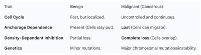
Comparison of Benign and Malignant Tumors
The following table summarizes key differences between benign and malignant tumors:
Trait | Benign | Malignant (Cancerous) |
|---|---|---|
Cell Cycle | Fast, but localized. | Uncontrolled and continuous. |
Anchorage Dependence | Present (Cells stay put). | Lost (Cells can migrate). |
Density-Dependent Inhibition | Partial loss. | Complete loss (Cells overlap). |
Genetics | Minor mutations. | Major chromosomal mutations/instability. |
Additional Info
Drug development often targets signal transduction pathways to treat diseases such as cancer.
Transcription factors are crucial in nuclear responses, regulating gene expression.
