 Back
BackLESSON 5 Cell Cycle and Tumor Formation: Structure, Regulation, and Cancer Development
Study Guide - Smart Notes
 Tailored notes based on your materials, expanded with key definitions, examples, and context.
Cell Cycle and Tumor Formation
Introduction
The cell cycle is a fundamental process in biology, governing how cells grow, replicate their DNA, and divide. Proper regulation of the cell cycle is essential for growth, development, and tissue repair. Disruption of cell cycle control can lead to uncontrolled cell proliferation and tumor formation, which underlies cancer development.
Organization of DNA in the Cell
Chromatin Structure
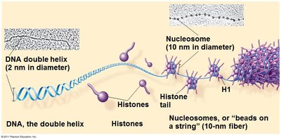
Chromatin is the complex of DNA and proteins (mainly histones) found in the nucleus of eukaryotic cells.
DNA wraps around histone proteins to form nucleosomes, which further coil and fold to form higher-order structures.
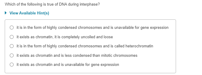
Chromatin exists in two main forms: euchromatin (less condensed, transcriptionally active) and heterochromatin (highly condensed, transcriptionally inactive).
Chromosomes and Chromatids
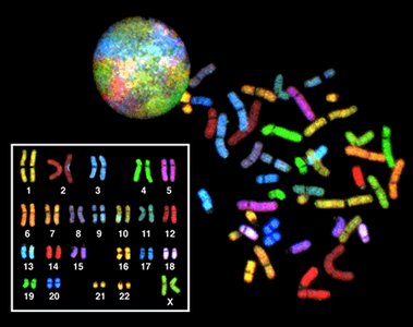
Chromosomes are highly condensed structures of chromatin visible during cell division.
Each duplicated chromosome consists of two sister chromatids joined at a region called the centromere.
Somatic cells are diploid (2N), containing two sets of chromosomes, while gametes are haploid (N).

The Cell Cycle: Phases and Regulation
Phases of the Cell Cycle
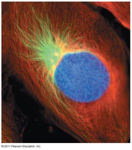
Interphase: The cell grows and DNA is replicated. It consists of three subphases:
G1 phase: Cell growth and normal functions
S phase: DNA synthesis (replication)
G2 phase: Preparation for mitosis
M phase (Mitotic phase): Includes mitosis (division of the nucleus) and cytokinesis (division of the cytoplasm).

Mitosis: Stages and Key Structures
Prophase: Chromatin condenses into visible chromosomes; mitotic spindle begins to form.
Prometaphase: Nuclear envelope breaks down; spindle fibers attach to kinetochores.
Metaphase: Chromosomes align at the metaphase plate.
Anaphase: Sister chromatids separate and move toward opposite poles.
Telophase: Nuclear envelopes reform; chromosomes decondense.
Cytokinesis: Cytoplasm divides, forming two daughter cells.
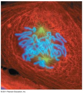
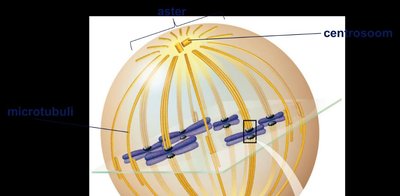
Mitotic Spindle and Microtubules
The mitotic spindle is a structure made of microtubules that segregates chromosomes during mitosis.
Microtubules are dynamic polymers of α- and β-tubulin that grow (polymerize) and shrink (depolymerize) as needed.
Kinetochores are protein complexes on chromosomes where spindle fibers attach.
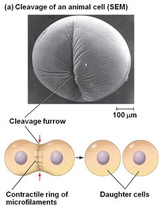
Cytokinesis
In animal cells, cytokinesis occurs via a cleavage furrow formed by a contractile ring of actin microfilaments and myosin.
Cell Renewal and Division in Human Tissues
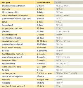
Cell Turnover Rates
Different tissues in the human body have varying rates of cell renewal, reflecting their function and exposure to damage.
Cell Type | Turnover Time |
|---|---|
Small intestine epithelium | 2-4 days |
Stomach | 2-9 days |
Blood Neutrophils | 1-5 days |
Blood platelets | 10 days |
Skin epidermal cells | 20-30 days |
Red blood cells (RBCs) | ~120 days |
Oocytes (female gametes) | Lifetime |
Cardiomyocytes | 0.5-1% per year |
Regulation of the Cell Cycle
Cell Cycle Control System
Progression through the cell cycle is regulated by internal and external signals.
Internal signals: Cyclins and cyclin-dependent kinases (Cdks) regulate transitions between phases.
External signals:
Growth factors: Chemical signals that stimulate cell division.
Anchorage dependence: Cells must be attached to a substrate to divide.
Density-dependent inhibition: Cells stop dividing when they become too crowded.
Loss of Cell Cycle Control and Tumor Formation
Transformation and Cancer
Transformation is the process by which a normal cell becomes a tumor cell, often due to mutations in genes controlling cell growth and division.
Key gene groups involved:
Proto-oncogenes: Normal genes that promote cell growth and division. When mutated, they become oncogenes that drive uncontrolled proliferation (e.g., ras).
Tumor-suppressor genes: Inhibit cell division, repair DNA, and promote apoptosis. Loss of function (e.g., p53) removes these brakes on the cell cycle.
Multistep Model of Cancer Development
Cancer typically arises from the accumulation of multiple mutations over time, affecting both proto-oncogenes and tumor-suppressor genes.
One mutation is usually insufficient; several genetic changes are required for malignant transformation.
Benign vs. Malignant Tumors
Benign tumors remain localized and do not invade other tissues.
Malignant tumors invade surrounding tissues and can spread (metastasize) via blood or lymphatic vessels, resulting in cancer.
Summary Table: Key Terms and Concepts
Term | Definition |
|---|---|
Chromatin | DNA-protein complex in the nucleus |
Chromosome | Condensed chromatin visible during cell division |
Sister chromatids | Identical copies of a chromosome joined at the centromere |
Interphase | Cell growth and DNA replication phase |
Mitosis | Nuclear division phase |
Cytokinesis | Cytoplasmic division phase |
Proto-oncogene | Gene promoting normal cell growth |
Oncogene | Mutated proto-oncogene causing cancer |
Tumor-suppressor gene | Gene inhibiting cell division |
Key Equations and Concepts
DNA replication during S phase:
Cell division produces two genetically identical daughter cells (except in meiosis).
Example: Skin Healing
When the skin is injured, cell division in the basal layer of the epidermis increases to replace lost or damaged cells, demonstrating the importance of regulated cell proliferation in tissue repair.
Additional info:
Mutations in ras (a proto-oncogene) can lead to constant cell division signals, while loss of p53 (a tumor-suppressor gene) impairs DNA repair and apoptosis, both contributing to cancer.
