 Back
BackCell Cycle, Mitosis, DNA Replication, and Protein Synthesis
Study Guide - Smart Notes
 Tailored notes based on your materials, expanded with key definitions, examples, and context.
Cell Cycle and Cell Division
Overview of the Cell Cycle
The cell cycle describes the sequence of events that a cell undergoes from its formation until it divides into two daughter cells. It consists of interphase (cell growth and DNA replication) and the mitotic phase (nuclear and cytoplasmic division).
Interphase: The period of cell growth and normal function, subdivided into G1, S, and G2 phases.
Mitotic (M) phase: The period where the cell divides its nucleus (mitosis) and cytoplasm (cytokinesis).
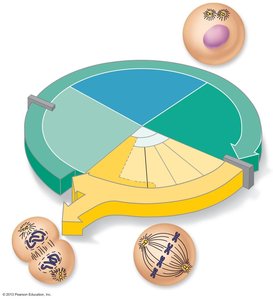
Phases of Interphase
G1 phase (Gap 1): Vigorous cell growth and metabolism. Cells that exit the cycle enter G0 phase, where they no longer divide.
S phase (Synthesis): DNA replication occurs, ensuring each daughter cell receives an identical set of chromosomes.
G2 phase (Gap 2): Final preparations for cell division, including synthesis of proteins required for mitosis.
Control of the Cell Cycle
Checkpoints: Critical control points (e.g., G1 and G2 checkpoints) ensure the cell is ready to proceed to the next phase.
Regulatory molecules: Cyclins and cyclin-dependent kinases (Cdks) regulate progression through the cell cycle.
Contact inhibition: Cells stop dividing when they come into contact with other cells.
DNA Replication
Mechanism of DNA Replication
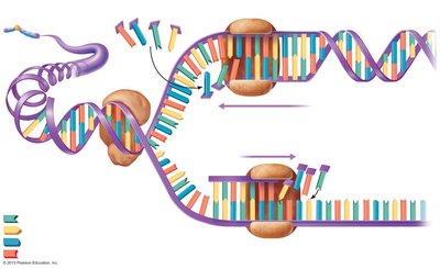
DNA replication is the process by which a cell copies its DNA before division. It is semiconservative, meaning each new DNA molecule consists of one old and one new strand.
Initiation: DNA helicase unwinds the double helix, forming replication bubbles with replication forks at each end.
Elongation: DNA polymerase adds complementary nucleotides to each template strand. The leading strand is synthesized continuously, while the lagging strand is synthesized in short fragments (Okazaki fragments) joined by DNA ligase.
Result: Two identical DNA molecules, each with one parental and one new strand.
Mitosis: Division of the Nucleus
Stages of Mitosis
Mitosis ensures equal distribution of replicated DNA to two daughter nuclei. It consists of four main stages:
Prophase: Chromosomes condense and become visible; centrosomes move to opposite poles; spindle fibers form; nuclear envelope breaks down.
Prometaphase/Late Prophase: Spindle fibers attach to kinetochores on chromosomes; nuclear envelope fragments completely.
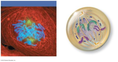
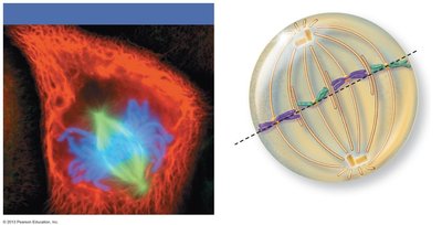
Metaphase: Chromosomes align at the metaphase plate (cell equator).
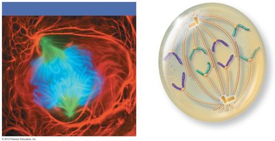
Anaphase: Sister chromatids separate at the centromere and are pulled toward opposite poles.
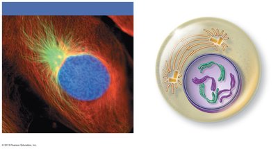
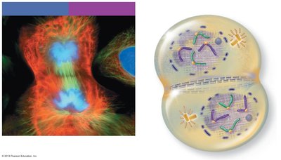
Telophase: Chromosomes decondense into chromatin; nuclear envelopes reform; nucleoli reappear; spindle apparatus disassembles.
Cytokinesis: Division of the cytoplasm, usually overlaps with telophase, resulting in two genetically identical daughter cells.
Protein Synthesis
From DNA to Protein: The Central Dogma
Protein synthesis involves two main steps: transcription (DNA to mRNA) and translation (mRNA to protein). The genetic code is based on triplets of nucleotides (codons) that specify amino acids.
Gene: A segment of DNA that codes for a specific polypeptide.
Triplet: Three DNA bases that code for one amino acid.
Codon: Three-base sequence on mRNA complementary to the DNA triplet.
Transcription: DNA to mRNA
Initiation: RNA polymerase binds to the promoter region of the gene with the help of transcription factors and unwinds the DNA.
Elongation: RNA polymerase synthesizes a complementary mRNA strand from the DNA template.
Termination: RNA polymerase stops transcription at a termination signal; the pre-mRNA is released.
Processing: Introns are removed, and exons are spliced together to form mature mRNA.
Translation: mRNA to Protein
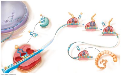
Initiation: The small ribosomal subunit binds to mRNA and the initiator tRNA (carrying methionine) at the start codon (AUG). The large subunit joins to form a functional ribosome.
Elongation: tRNAs bring amino acids to the ribosome, matching their anticodons to mRNA codons. Peptide bonds form between amino acids, and the ribosome moves along the mRNA.
Termination: When a stop codon (UAA, UAG, UGA) is reached, a release factor binds, releasing the completed polypeptide and disassembling the ribosome.
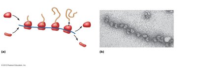
Polyribosomes: Multiple ribosomes can translate a single mRNA simultaneously, producing many copies of a protein.
Role of the Rough Endoplasmic Reticulum (ER) in Protein Synthesis
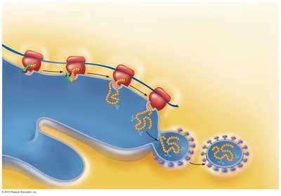
Proteins destined for secretion or membrane insertion are synthesized on ribosomes bound to the rough ER.
A signal-recognition particle (SRP) directs the ribosome-mRNA complex to the ER membrane, where the growing polypeptide enters the ER lumen.
Proteins are modified (e.g., glycosylation) and folded, then packaged into vesicles for transport to the Golgi apparatus.
Summary Table: Key Steps in the Flow of Genetic Information
Step | Location | Main Molecules Involved | Key Events |
|---|---|---|---|
DNA Replication | Nucleus | DNA, DNA polymerase, ligase | Semiconservative synthesis of new DNA strands |
Transcription | Nucleus | DNA, RNA polymerase, mRNA | DNA sequence copied into mRNA |
RNA Processing | Nucleus | pre-mRNA, spliceosome | Introns removed, exons joined to form mature mRNA |
Translation | Cytoplasm (ribosome) | mRNA, tRNA, rRNA, amino acids | mRNA codons translated into amino acid sequence |
Additional info:
Cell division is tightly regulated by internal and external signals to prevent uncontrolled proliferation (cancer).
Protein synthesis is fundamental for cell structure and function, as proteins serve as enzymes, structural components, and signaling molecules.
