 Back
BackCell Division and Reproduction: Mitosis, Meiosis, and Chromosomal Alterations
Study Guide - Smart Notes
 Tailored notes based on your materials, expanded with key definitions, examples, and context.
Cell Division and Reproduction
Asexual and Sexual Reproduction
Cell division is fundamental for the continuity of life, enabling organisms to reproduce, grow, repair tissues, and maintain their genetic integrity. There are two main types of reproduction:
Asexual reproduction: Produces offspring genetically identical to the parent, involving only one parent. Common in single-celled organisms and for growth and repair in multicellular organisms.
Sexual reproduction: Produces genetically varied offspring by combining genes from two parents, involving the formation of gametes (sperm and egg).
Prokaryotic Cell Division: Binary Fission
Mechanism of Binary Fission
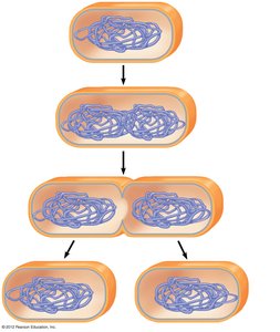
Prokaryotes (bacteria and archaea) reproduce by binary fission, a process that results in two genetically identical daughter cells. The prokaryotic chromosome is a single, circular DNA molecule.
Step 1: Chromosome duplication and separation of copies
Step 2: Cell elongation and movement of chromosome copies
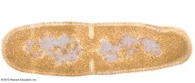
Step 3: Division into two daughter cells
Example: Escherichia coli dividing by binary fission.
Eukaryotic Cell Cycle and Mitosis
Structure of Eukaryotic Chromosomes
Eukaryotic cells are more complex, containing multiple linear chromosomes within a nucleus. Chromosomes are composed of chromatin (DNA and proteins).
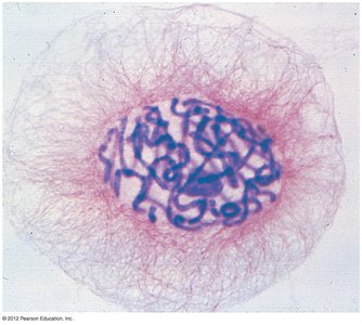
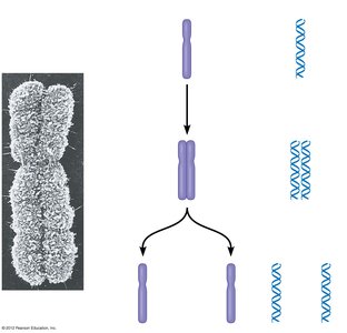
Before division, chromosomes duplicate, forming sister chromatids joined at a centromere.
During cell division, sister chromatids separate, ensuring each daughter cell receives an identical set of chromosomes.
The Cell Cycle
The cell cycle is an ordered sequence of events from the formation of a cell to its own division. It consists of:
Interphase: Cell growth and chromosome duplication (G1, S, G2 phases)
Mitotic (M) phase: Division of the nucleus (mitosis) and cytoplasm (cytokinesis)
Phases of Mitosis
Mitosis is divided into several stages:
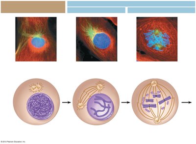
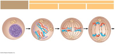
Prophase: Chromosomes condense, spindle forms
Prometaphase: Nuclear envelope breaks down, spindle fibers attach to kinetochores
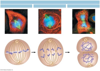
Metaphase: Chromosomes align at the cell equator
Anaphase: Sister chromatids separate and move to opposite poles
Telophase: Nuclear envelopes reform, chromosomes decondense
Cytokinesis
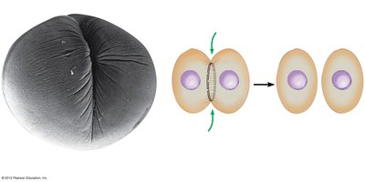
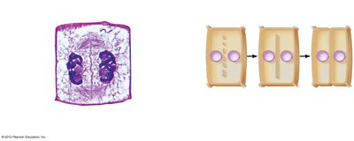
Cytokinesis divides the cytoplasm, resulting in two daughter cells. The process differs between animal and plant cells:
Animal cells: Formation of a cleavage furrow by a contractile ring of microfilaments.
Plant cells: Formation of a cell plate from vesicles, which develops into a new cell wall.
Functions of Mitosis
Growth of multicellular organisms
Repair and replacement of damaged cells
Asexual reproduction in some organisms
Meiosis and Sexual Life Cycles
Homologous Chromosomes and Chromosome Number
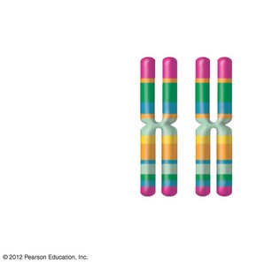
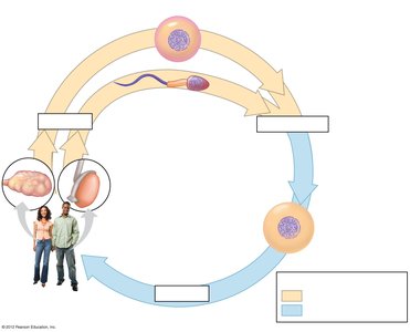
Somatic cells are diploid (2n), containing pairs of homologous chromosomes. Gametes are haploid (n), containing one set of chromosomes.
Overview of Meiosis
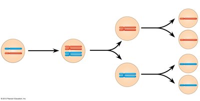
Meiosis reduces the chromosome number by half, producing four genetically unique haploid gametes from a diploid cell. It consists of two divisions: meiosis I and meiosis II.
Phases of Meiosis
Meiosis I: Homologous chromosomes separate
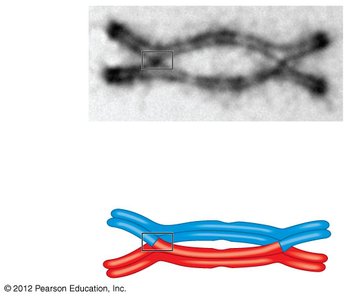
Prophase I: Synapsis and crossing over form tetrads
Metaphase I: Tetrads align at the equator
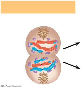
Anaphase I: Homologous chromosomes separate
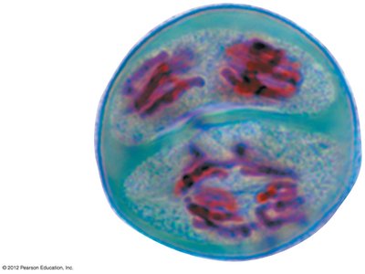
Telophase I and Cytokinesis: Two haploid cells form
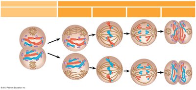
Meiosis II: Sister chromatids separate (similar to mitosis)
Prophase II, Metaphase II, Anaphase II, Telophase II, and Cytokinesis
Comparison of Mitosis and Meiosis
Mitosis: Produces two genetically identical diploid cells for growth and repair.
Meiosis: Produces four genetically unique haploid gametes for sexual reproduction.
Genetic Variation in Sexual Reproduction
Sources of Genetic Variation
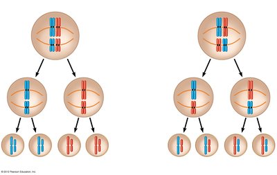
Independent assortment: Random orientation of homologous pairs during metaphase I creates many possible combinations (2n where n = haploid number).
Random fertilization: Any sperm can fertilize any egg, increasing variability.
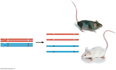
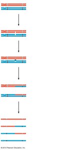
Crossing over: Exchange of genetic material between nonsister chromatids during prophase I produces recombinant chromosomes.
Alterations of Chromosome Number and Structure
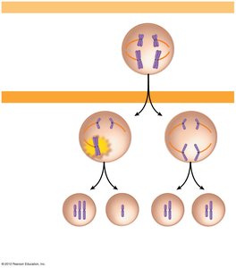
Nondisjunction
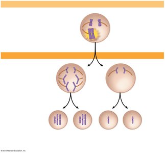
Nondisjunction is the failure of chromosomes or chromatids to separate properly during meiosis, resulting in gametes with abnormal chromosome numbers. Fertilization involving such gametes leads to aneuploidy (e.g., trisomy or monosomy).
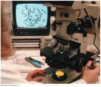
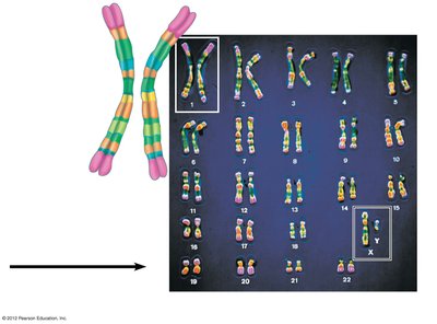
Karyotyping
A karyotype is an ordered display of an individual's chromosomes, used to detect chromosomal abnormalities.
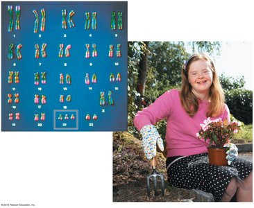
Down Syndrome (Trisomy 21)
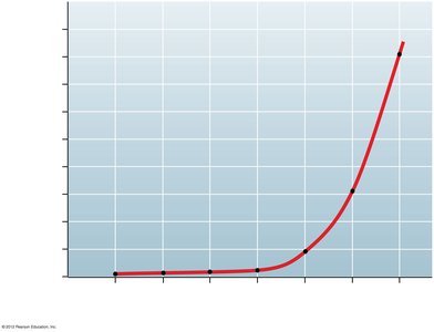
Down syndrome is caused by an extra copy of chromosome 21. Symptoms include intellectual disability, characteristic facial features, heart defects, and increased risk of certain diseases. The risk increases with maternal age.
Sex Chromosome Abnormalities
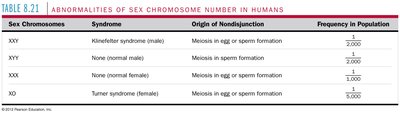
Abnormal numbers of sex chromosomes (e.g., XXY, XYY, XXX, XO) usually have less severe effects due to X-inactivation and the small size of the Y chromosome.
Sex Chromosomes | Syndrome | Origin of Nondisjunction | Frequency in Population |
|---|---|---|---|
XXY | Klinefelter syndrome (male) | Meiosis in egg or sperm formation | 1/2,000 |
XYY | None (normal male) | Meiosis in sperm formation | 1/2,000 |
XXX | None (normal female) | Meiosis in egg or sperm formation | 1/1,000 |
XO | Turner syndrome (female) | Meiosis in egg or sperm formation | 1/5,000 |
Polyploidy and Chromosomal Rearrangements
Polyploidy: Organisms with more than two sets of chromosomes, common in plants.
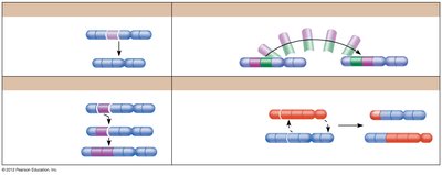
Chromosomal rearrangements: Deletions, duplications, inversions, and translocations can cause genetic disorders or cancer.
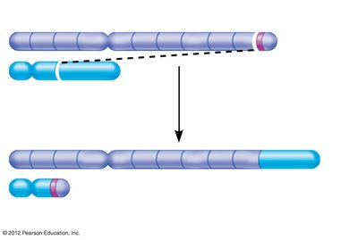
Example: Chronic myelogenous leukemia (CML) results from a reciprocal translocation between chromosomes 9 and 22, creating the "Philadelphia chromosome." Cancer is usually not inherited because it often arises from somatic cell mutations.
