 Back
BackCell Division and the Cell Cycle: Structure, Function, and Mechanisms
Study Guide - Smart Notes
 Tailored notes based on your materials, expanded with key definitions, examples, and context.
Cell Division: Importance and Overview
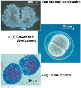
Why Care About Cell Division?
Cell division is a fundamental biological process essential for growth, development, tissue renewal, and reproduction in all living organisms. It ensures the continuity of life by producing new cells from pre-existing ones, as famously stated by Rudolf Virchow: "Omnis cellula e cellula" (every cell from a cell).
Growth and Development: Multicellular organisms grow by increasing their cell number through division.
Tissue Renewal: Damaged or dead cells are replaced via cell division, maintaining tissue health.
Asexual Reproduction: Many unicellular organisms reproduce by dividing into two identical cells.
Mechanisms of Cell Division
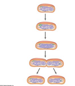
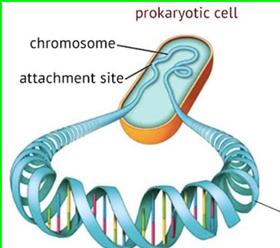
Binary Fission in Prokaryotes
Prokaryotic cells, such as bacteria, divide by a process called binary fission. This is a simpler mechanism compared to eukaryotic cell division and involves the replication of the single, circular chromosome followed by division of the cytoplasm.
Steps: DNA replication, chromosome segregation, and cytokinesis.
Result: Two genetically identical daughter cells.
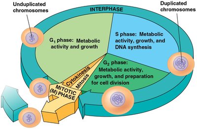
Cell Cycle in Eukaryotes
The cell cycle describes the ordered sequence of events that a eukaryotic cell undergoes from its formation to its own division. It consists of interphase (cell growth and DNA replication) and the mitotic (M) phase (mitosis and cytokinesis).
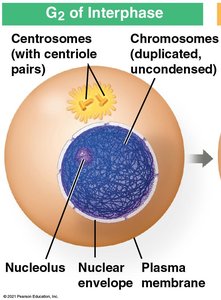
Interphase: Includes G1 (growth), S (DNA synthesis), and G2 (preparation for division).
M Phase: Includes mitosis (nuclear division) and cytokinesis (cytoplasmic division).
DNA Replication and Packaging
When and How is DNA Replicated?
DNA replication occurs during the S phase of interphase. Accurate replication is essential to ensure that each daughter cell receives an identical set of genetic information.
Timing: S phase of interphase.
Purpose: To duplicate the cell's genetic material before division.
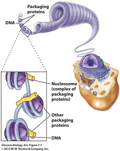
DNA Packaging
Before cell division, DNA is tightly packaged into structures called chromosomes. This packaging involves winding DNA around proteins called histones, forming nucleosomes and higher-order structures.
Chromosome Structure and Terminology
Key Terms
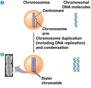
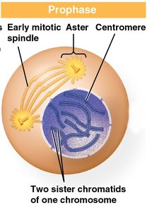
Chromosome: A DNA molecule with associated proteins, visible during cell division.
Chromatin: The less condensed form of DNA and protein found in the nucleus during interphase.
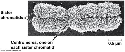
Sister Chromatids: Two identical copies of a chromosome, joined at the centromere, formed after DNA replication.
Centromere: The region where sister chromatids are most closely attached.
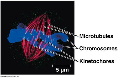
Kinetochore: A protein complex on the centromere where spindle fibers attach during mitosis.
The Stages of Mitosis
Overview of Mitosis
Mitosis is the process by which a eukaryotic cell separates its duplicated chromosomes into two identical nuclei. It is divided into several stages, each with distinct events:
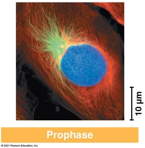
Prophase: Chromosomes condense, spindle apparatus forms, nuclear envelope begins to break down.
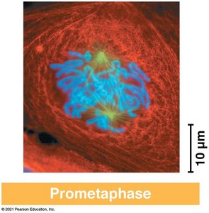
Prometaphase: Nuclear envelope fragments, spindle fibers attach to kinetochores.
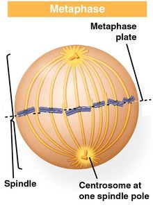
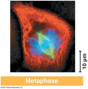
Metaphase: Chromosomes align at the metaphase plate.
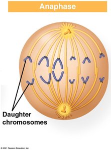
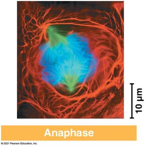
Anaphase: Sister chromatids separate and move toward opposite poles.
Telophase: Nuclear envelopes reform, chromosomes decondense.
Prophase
Chromosomes condense and become visible.
Mitotic spindle begins to form.
Prometaphase
Nuclear envelope fragments.
Spindle microtubules attach to kinetochores.
Metaphase
Chromosomes align at the metaphase plate (cell equator).
Spindle fibers attach to kinetochores of each chromosome.
Anaphase
Sister chromatids separate at the centromere.
Chromatids (now daughter chromosomes) move to opposite poles.
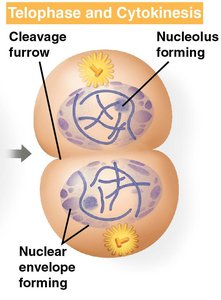
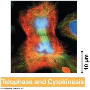
Telophase and Cytokinesis
Nuclear envelopes reform around chromosomes.
Chromosomes decondense.
Cytokinesis divides the cytoplasm, forming two daughter cells.
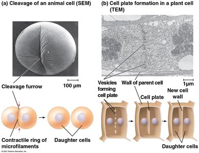
Cytokinesis: Animal vs. Plant Cells
Mechanisms of Cytoplasmic Division
Animal Cells: Cytokinesis occurs by cleavage, forming a cleavage furrow that pinches the cell in two.
Plant Cells: Cytokinesis occurs by cell plate formation, where vesicles coalesce at the center to form a new cell wall.
Microscopic Observation of Mitosis
Identifying Stages in Real Cells
Microscopic examination of dividing cells (e.g., onion root tip) reveals cells at various stages of mitosis, allowing for the identification of interphase, prophase, metaphase, anaphase, and telophase.
Summary Table: Key Terms and Concepts
Term | Definition |
|---|---|
Chromosome | Condensed DNA molecule visible during cell division |
Chromatin | Uncondensed DNA-protein complex in interphase |
Sister Chromatid | One of two identical halves of a duplicated chromosome |
Centromere | Region where sister chromatids are joined |
Kinetochore | Protein structure on centromere for spindle attachment |
Cytokinesis | Division of the cytoplasm into two cells |
Binary Fission | Prokaryotic cell division mechanism |
Additional info:
The cell cycle is tightly regulated by checkpoints to ensure accurate division.
Errors in cell division can lead to diseases such as cancer.
