Back
Cell Division, Mendelian Genetics, and Human Genetic Analysis: Study Guide
Study Guide - Smart Notes
 Tailored notes based on your materials, expanded with key definitions, examples, and context.
Cell Division
Somatic vs. Germ Cells
In eukaryotic organisms, cells are classified as somatic cells or germ cells based on their function and role in the organism.
Somatic Cells: Body cells responsible for growth, repair, and maintenance. They are diploid (2n) and divide by mitosis.
Germ Cells: Specialized cells that give rise to gametes (sperm and eggs) through meiosis. They are involved in sexual reproduction and produce haploid (n) cells.
Understanding these distinctions is essential for grasping how organisms grow and reproduce.
Mitosis vs. Meiosis
Mitosis and meiosis are two types of nuclear division with distinct purposes:
Mitosis: One division cycle; produces two genetically identical diploid cells for growth and repair.
Meiosis: Two division cycles; produces four genetically unique haploid gametes for sexual reproduction.
Chromosome Behavior: In mitosis, chromosomes line up individually; in meiosis, homologous chromosomes pair up and undergo crossing over, increasing genetic variation.
Chromatin, Chromatids, and Chromosomes
Chromatin: DNA-protein complex in non-dividing cells; long, thin fibers.
Chromosomes: Condensed chromatin visible during cell division; each contains one DNA molecule.
Chromatids: Duplicated halves of a chromosome, called sister chromatids, joined at the centromere before separation.
Haploid vs. Diploid Cells
Diploid (2n): Two sets of homologous chromosomes (e.g., human somatic cells: 46 chromosomes).
Haploid (n): One set of chromosomes (e.g., human gametes: 23 chromosomes).
Fertilization restores the diploid state, ensuring genetic continuity across generations.
Chromosome Condensation
Chromosome condensation is essential for the accurate and equal distribution of genetic material during cell division. Condensed chromosomes are easier to segregate, ensuring each daughter cell receives the correct genetic information and maintaining genetic stability.
Eukaryotic Cell Cycle
Phases of the Cell Cycle
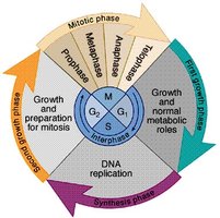
The eukaryotic cell cycle consists of interphase (G1, S, G2) and the mitotic phase (M), followed by cytokinesis.
G1 Phase: Cell growth and normal metabolic roles.
S Phase: DNA replication.
G2 Phase: Preparation for mitosis.
Mitosis (M Phase): Division of the nucleus.
Cytokinesis: Division of the cytoplasm, forming two daughter cells.
Mitosis
Stages of Mitosis
Mitosis is divided into several stages, each with specific activities and roles for the cytoskeleton and organelles:
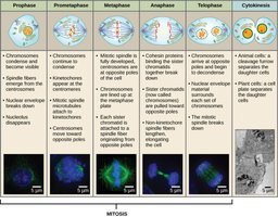
Prophase: Chromosomes condense; mitotic spindle forms; nuclear envelope breaks down.
Metaphase: Chromosomes align at the metaphase plate; spindle fibers attach to centromeres.
Anaphase: Sister chromatids separate and move to opposite poles.
Telophase: Chromatids reach poles; nuclear envelopes reform; chromosomes decondense.
Cytokinesis: Cytoplasm divides, resulting in two identical daughter cells.
Meiosis
Haploid and Diploid; Sets of Chromosomes
Diploid (2n): Two sets of homologous chromosomes (e.g., human somatic cells: 46 chromosomes).
Haploid (n): One set of chromosomes (e.g., human gametes: 23 chromosomes).
Homologous chromosomes are pairs, one from each parent, carrying genes for the same traits.
Homologous, Maternal, and Paternal Chromosomes
Homologous Chromosomes: Pairs with the same structure and gene loci, but possibly different alleles.
Maternal Chromosome: Inherited from the mother.
Paternal Chromosome: Inherited from the father.
Comparison of Mitosis and Meiosis
Mitosis: One division, two identical diploid cells, no genetic variation.
Meiosis: Two divisions, four unique haploid cells, genetic variation via crossing over and independent assortment.
Meiosis I and II: Stages and Activities
Meiosis consists of two sequential divisions:
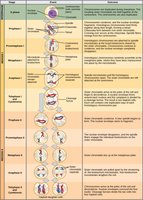
Meiosis I: Homologous chromosomes pair, cross over, and separate into two haploid cells.
Meiosis II: Sister chromatids separate, resulting in four haploid cells.
Mendelian Genetics
Basic Terms in Genetics
Dominant: Allele expressed in the phenotype when present.
Recessive: Allele expressed only when two copies are present.
Homologous Chromosomes: Chromosome pairs with the same structure and gene loci.
Homozygous: Two identical alleles for a gene.
Heterozygous: Two different alleles for a gene.
Genotype: Genetic makeup of an organism.
Phenotype: Observable traits of an organism.
Allele: Different versions of a gene.
Monohybrid Crosses
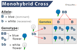
A monohybrid cross examines the inheritance of a single trait. Punnett squares are used to predict genotype and phenotype ratios.
Testcross (Back Cross)
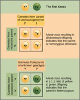
A testcross determines the genotype of an individual with a dominant phenotype by crossing it with a homozygous recessive individual. The offspring's phenotypes reveal the unknown genotype.
Variations on the Mendelian Theme
Complete Dominance
In complete dominance, the heterozygote displays the same phenotype as the homozygous dominant. Example: Mendel's pea plants (purple flowers dominant over white).
Incomplete Dominance
In incomplete dominance, the heterozygote has an intermediate phenotype. Example: Red and white snapdragons produce pink offspring.
Codominance and Multiple Alleles (ABO Blood Types)
Codominance occurs when both alleles are fully expressed in the heterozygote. The ABO blood group system is an example, with three alleles (IA, IB, i) producing four blood types (A, B, AB, O).
Pleiotropy
Pleiotropy is when one gene affects multiple traits. Example: Sickle-cell disease affects red blood cell shape, anemia, and organ function.
Polygenic Inheritance
Polygenic inheritance involves multiple genes influencing a single trait, resulting in continuous variation. Examples: human height, skin color.
Epistasis (Labrador Coat Color)
Epistasis occurs when one gene affects the expression of another. In Labradors, two genes interact to determine coat color (black, chocolate, yellow).
Environmental Effects
Both genetics and environment shape an organism. External factors (temperature, light, diet) and internal factors (hormones) can influence gene expression and phenotype.
Chromosomes and Human Genetics
Sex Chromosomes and Autosomes
Sex Chromosomes: X and Y chromosomes determine biological sex (XX = female, XY = male).
Autosomes: Non-sex chromosomes; humans have 22 pairs.
X-linked Inheritance
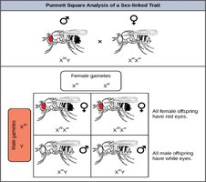
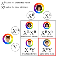
X-linked traits are inherited via the X chromosome. Males are more likely to express X-linked recessive traits (e.g., colorblindness, hemophilia) because they have only one X chromosome.
Linkage
Linkage refers to genes located close together on the same chromosome, which tend to be inherited together. Linkage maps show the relative positions of genes based on recombination frequencies.
Chromosome Mutations
Deletion: Loss of a chromosome segment (e.g., cri du chat syndrome).
Duplication: Repetition of a chromosome segment.
Inversion: Reversal of a chromosome segment.
Translocation: Segment moves to a nonhomologous chromosome (e.g., CML).
Human Genetic Analysis
Pedigree Analysis
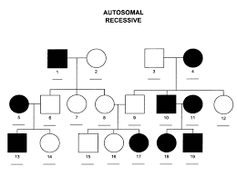
Pedigrees are diagrams that track inheritance patterns across generations. Symbols: circles (females), squares (males), shaded (affected), unshaded (unaffected). Pedigrees help determine modes of inheritance (autosomal dominant, autosomal recessive, X-linked, Y-linked).
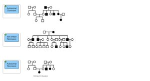
Single Gene Inheritance Patterns
Autosomal Recessive: Albinism, sickle-cell anemia, Tay-Sachs, cystic fibrosis, PKU.
Autosomal Dominant: Huntington’s disease, polydactyly, familial hypercholesterolemia.
X-linked Recessive: Colorblindness, hemophilia.
X-linked Dominant: Faulty dental enamel.
Y-linked: SRY gene, sperm production, hair in the ears.