 Back
BackCell Division: Mitosis, Cell Cycle, and Cancer
Study Guide - Smart Notes
 Tailored notes based on your materials, expanded with key definitions, examples, and context.
How Cells Divide
Overview of Cell Division
Cell division is a fundamental process by which cells reproduce, enabling growth, development, and tissue repair in multicellular organisms. In prokaryotes, division occurs by binary fission, while eukaryotes undergo mitosis and cytokinesis. Proper regulation of cell division is essential for maintaining genetic stability and preventing diseases such as cancer.
Bacterial Cell Division: Binary Fission
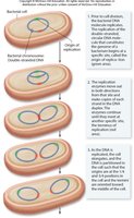
Mechanism of Binary Fission
Binary fission is the process by which bacteria reproduce asexually, producing two genetically identical daughter cells.
Bacteria possess a single, circular chromosome that is replicated prior to division.
Replication begins at the origin of replication and proceeds bidirectionally until the entire chromosome is copied.
New chromosomes are segregated to opposite ends of the cell.
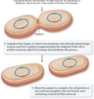
A septum forms, dividing the cell into two.
Eukaryotic Chromosomes and Karyotype
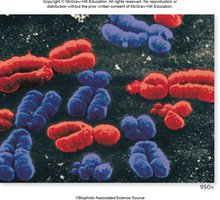
Chromosome Structure and Number
Humans have 46 chromosomes arranged in 23 pairs.
Each chromosome consists of DNA and associated proteins, forming a compact structure visible during cell division.
Abnormal chromosome numbers can lead to genetic disorders.
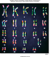
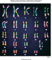
Karyotype
A karyotype is the complete set of chromosomes in an organism, arranged by size, centromere position, and staining pattern.
Humans are diploid (2n), possessing two sets of chromosomes (one from each parent).
Haploid (n) cells, such as gametes, contain a single set of chromosomes.
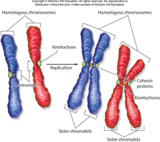
Homologous chromosomes are pairs with the same genes but possibly different alleles.
DNA Replication and Chromosome Structure
DNA Replication
Before replication, each chromosome contains a single DNA molecule.
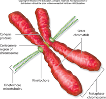
After replication, each chromosome consists of two identical sister chromatids joined at the centromere by cohesin proteins.
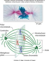
Centromere and Kinetochore
The centromere is the constricted region of the chromosome where sister chromatids are attached.
The kinetochore is a protein complex at the centromere that serves as the attachment site for spindle microtubules during mitosis.
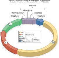
The Eukaryotic Cell Cycle
Phases of the Cell Cycle
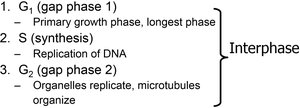
The eukaryotic cell cycle consists of interphase (G1, S, G2) and the M phase (mitosis and cytokinesis). The duration of the cell cycle varies by cell type and organism.
G1 phase (Gap 1): Primary growth phase; cell increases in size and prepares for DNA replication.
S phase (Synthesis): DNA is replicated.
G2 phase (Gap 2): Organelles replicate, and microtubules organize in preparation for mitosis.
M phase: Includes mitosis (nuclear division) and cytokinesis (cytoplasmic division).
G0 phase: Resting phase where cells are not actively dividing.
Mitosis: Phases and Events
Overview of Mitosis
Mitosis is the process by which a eukaryotic cell divides its nucleus and distributes duplicated chromosomes into two daughter nuclei. It is divided into five phases: prophase, prometaphase, metaphase, anaphase, and telophase.
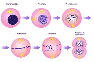
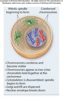
Prophase
Chromosomes condense and become visible under a light microscope.
The spindle apparatus begins to form as centrioles move to opposite poles (in animal cells).
Asters (radial microtubule arrays) form in animal cells.
The nuclear envelope breaks down.
Prometaphase
Microtubules attach to chromosomes at the kinetochore.
Chromosomes begin moving toward the metaphase plate.
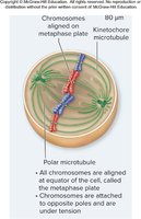
Metaphase
Chromosomes align along the metaphase plate (an imaginary plane at the cell's center).
This alignment ensures each daughter cell will receive one copy of each chromosome.
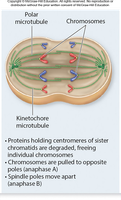
Anaphase
Centromeres split, separating sister chromatids.
Cohesin proteins are removed, and microtubules pull chromatids to opposite poles.
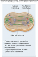
Telophase
The spindle apparatus disassembles.
Nuclear envelopes reform around each set of chromosomes.
Chromosomes begin to uncoil, and nucleoli reappear.
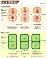
Cytokinesis
Division of the Cytoplasm
Cytokinesis is the process of dividing the cytoplasm to form two separate daughter cells.
In animal cells, a cleavage furrow forms via constriction of actin filaments.
In plant cells, a cell plate forms between the nuclei.
In fungi and some protists, mitosis occurs within the nucleus, and division is coupled with cytokinesis.
Control of the Cell Cycle
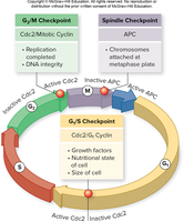
Checkpoints and Regulation
The cell cycle is regulated at specific checkpoints to ensure accuracy and respond to internal/external signals.
G1/S checkpoint: Cell commits to division; influenced by external signals.
G2/M checkpoint: Cell checks for successful DNA replication before mitosis.
Spindle checkpoint (late metaphase): Ensures all chromosomes are attached to the spindle before anaphase.
Growth Factors
Growth factors are signaling molecules that stimulate cell division by activating intracellular pathways.
Example: Platelet-derived growth factor (PDGF) activates a MAP kinase cascade via receptor tyrosine kinases.
Cancer and Cell Cycle Control
Uncontrolled Cell Division
Cancer results from unrestrained, uncontrolled cell growth due to failure of cell cycle regulation.
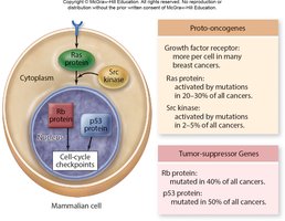
Two main gene types involved: proto-oncogenes and tumor-suppressor genes.
Proto-oncogenes
Normal genes that promote cell division; mutations convert them to oncogenes that drive cancer.
Examples include genes encoding growth factor receptors and signal transduction proteins.
Only one mutated copy is needed for uncontrolled division.
Tumor-suppressor Genes
Genes that inhibit cell division or promote apoptosis.
Both copies must be inactivated for cancer to develop.
Examples: p53 gene (mutated in ~50% of cancers), Rb gene (retinoblastoma susceptibility).
Summary Table: Key Differences in Cell Division
Process | Organism | Key Features |
|---|---|---|
Binary Fission | Bacteria (Prokaryotes) | Single circular chromosome, no mitosis, septum formation |
Mitosis | Eukaryotes | Multiple linear chromosomes, spindle apparatus, five phases |
Key Terms
Chromatid: One of two identical halves of a replicated chromosome.
Centromere: Region where sister chromatids are joined.
Kinetochore: Protein structure on centromere for spindle attachment.
Cohesin: Protein complex holding sister chromatids together.
Metaphase Plate: Imaginary plane where chromosomes align during metaphase.
Equations and Additional Info
Diploid number: (humans)
Haploid number: (humans)
