 Back
BackCell Division: Mitosis, Meiosis, and Cancer – A Comprehensive Study Guide
Study Guide - Smart Notes
 Tailored notes based on your materials, expanded with key definitions, examples, and context.
Cell Division: An Overview
Why Do Cells Divide?
Cell division is a fundamental process in all living organisms, essential for reproduction, growth, development, and tissue repair. The mechanisms and outcomes of cell division differ between prokaryotes and eukaryotes, and between somatic and reproductive cells.
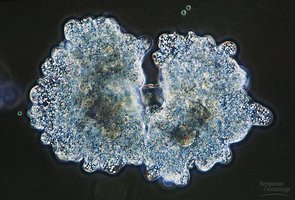
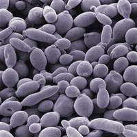
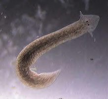
Asexual Reproduction: Involves the production of new individuals without the fusion of gametes. Offspring are genetically identical to the parent (clones). Examples include budding in yeast, fragmentation in lichens, and fission in planaria.
Growth and Development: Multicellular organisms grow from a single fertilized egg (zygote) to an adult through repeated cell divisions.
Repair and Maintenance: Cell division replaces damaged or dead cells, maintaining tissue integrity.
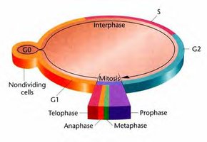
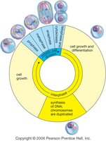
The Cell Cycle
Phases of the Cell Cycle
The cell cycle is a regulated series of events that cells undergo as they grow and divide. It consists of interphase (G1, S, G2), mitosis (M phase), and cytokinesis. Some cells may exit the cycle into a quiescent state (G0).
G1 Phase: Cell grows and carries out normal functions.
S Phase: DNA is replicated.
G2 Phase: Further growth and preparation for mitosis.
Mitosis (M Phase): Division of the nucleus.
Cytokinesis: Division of the cytoplasm, resulting in two daughter cells.
G0 Phase: Non-dividing state; some specialized cells (e.g., neurons, muscle cells) remain here permanently.
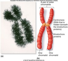
Chromosomes and Genome Organization
Chromosomes are the structural units of genetic material. Eukaryotic cells have multiple linear chromosomes, while prokaryotes typically have a single circular chromosome. Chromatin is the complex of DNA and proteins that forms chromosomes.
Sister Chromatids: Identical copies of a chromosome, joined at the centromere, produced during DNA replication.
Centromere: The region where sister chromatids are attached.
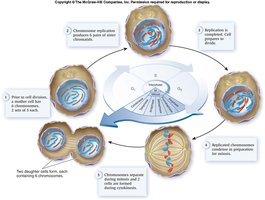
Mitosis
Phases of Mitosis
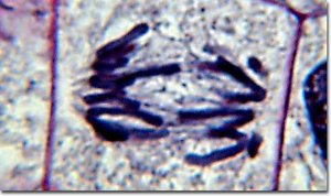
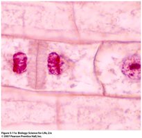
Mitosis is the process by which somatic cells divide to produce two genetically identical daughter cells. It consists of four main phases: prophase, metaphase, anaphase, and telophase (PMAT).
Prophase: Chromatin condenses into visible chromosomes, the nuclear envelope breaks down, and the mitotic spindle forms. In animal cells, centrioles migrate to opposite poles; plant cells lack centrioles.
Metaphase: Chromosomes align at the metaphase plate (equatorial plane). Spindle fibers attach to centromeres.
Anaphase: Sister chromatids separate and move toward opposite poles, now called daughter chromosomes.
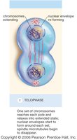
Telophase: Chromosomes decondense, nuclear envelopes reform, and cytokinesis begins.
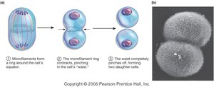
Cytokinesis
Cytokinesis is the division of the cytoplasm, resulting in two separate daughter cells. The process differs between animal and plant cells:
Animal Cells: A cleavage furrow forms, pinching the cell membrane until the cell splits.
Plant Cells: A cell plate forms, eventually developing into a new cell wall that separates the daughter cells.
Binary Fission in Prokaryotes
Prokaryotic cells divide by binary fission, a simpler process than mitosis:
The single circular chromosome replicates.
Origins of replication move to opposite ends of the cell.
The cell elongates, and the plasma membrane pinches inward.
The cell splits, producing two genetically identical cells.
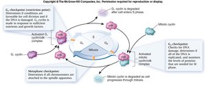
Regulation of the Cell Cycle
Cell Cycle Checkpoints
Checkpoints are control mechanisms that ensure the fidelity of cell division. Cells must pass these checkpoints to proceed through the cycle:
G1/S Checkpoint: Checks for cell size, nutrients, and DNA integrity.
G2/M Checkpoint: Ensures DNA replication is complete and checks for DNA damage.
Metaphase/Anaphase Checkpoint: Ensures all chromosomes are properly attached to the spindle apparatus.
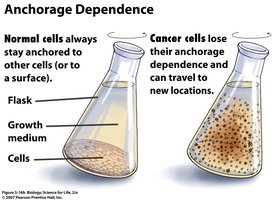
Density-Dependent Inhibition and Anchorage Dependence
Normal cells stop dividing when they become crowded (density-dependent inhibition) and require attachment to a substrate (anchorage dependence) to divide. Cancer cells often lose these controls.
Cancer and Cell Division
Loss of Cell Cycle Control
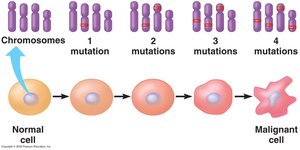
Cancer results from the loss of normal cell cycle regulation, leading to uncontrolled cell division. Carcinogens (e.g., chemicals, UV light, viruses) can cause mutations that disrupt checkpoint controls.
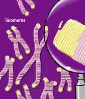
Telomeres: Repetitive DNA sequences at chromosome ends that shorten with each division. Normal cells stop dividing when telomeres are depleted; cancer cells often activate telomerase to maintain telomere length.
Hayflick Limit: The maximum number of times a normal human cell can divide (about 50 times).
Multi-hit Hypothesis: Multiple mutations are typically required for a cell to become cancerous.
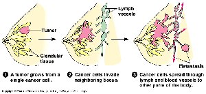
Types of Tumors
Benign Tumor: Cells proliferate abnormally but do not invade other tissues.
Malignant Tumor: Cancer cells invade surrounding tissues and can spread (metastasize) to distant organs, often via the lymphatic system.
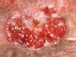
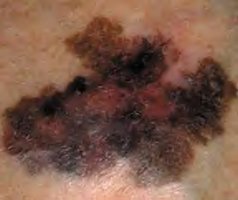
Skin Cancer and Sunlight
Ultraviolet (UV) light can damage DNA, leading to skin cancer. Types include:
Basal Cell Carcinoma: Most common, least malignant.
Squamous Cell Carcinoma: Grows rapidly, may metastasize.
Malignant Melanoma: Most dangerous, highly metastatic.
Meiosis: The Formation of Sex Cells
Overview of Meiosis
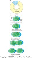
Meiosis is a specialized form of cell division that reduces the chromosome number by half, producing four genetically unique haploid cells (gametes). It consists of two consecutive divisions: meiosis I and meiosis II.
Haploid (n): Cells with one set of chromosomes (e.g., gametes).
Diploid (2n): Cells with two sets of chromosomes (e.g., somatic cells).
Genetic Variation in Meiosis
Crossing Over: Exchange of genetic material between homologous chromosomes during prophase I, increasing genetic diversity.
Random Alignment: Homologous chromosomes align randomly at the metaphase plate during metaphase I, resulting in numerous possible combinations of chromosomes in gametes.
Stages of Meiosis
Meiosis I: Homologous chromosomes separate, reducing chromosome number by half.
Meiosis II: Sister chromatids separate, similar to mitosis.
Mitosis vs. Meiosis
Feature | Mitosis | Meiosis |
|---|---|---|
Number of Divisions | 1 | 2 |
Number of Daughter Cells | 2 | 4 |
Genetic Identity | Identical | Unique |
Chromosome Number | Diploid (2n) | Haploid (n) |
Role | Growth, repair | Gamete production |
Gametogenesis
Spermatogenesis
Spermatogenesis is the process of sperm formation in males. It results in four functional, haploid sperm cells from each primary spermatocyte.
Oogenesis
Oogenesis is the process of egg formation in females. It produces one functional ovum and three polar bodies (which degenerate) from each primary oocyte.
Comparison of Spermatogenesis and Oogenesis
Feature | Spermatogenesis | Oogenesis |
|---|---|---|
Number of Functional Gametes | 4 | 1 |
Timing | Continuous after puberty | Begins before birth, completes after fertilization |
Polar Bodies | None | 3 (degenerate) |
Sexual Reproduction and Life Cycles
Sexual reproduction involves the fusion of two haploid gametes from different individuals, resulting in offspring with genetic variation. This variation is crucial for evolution and adaptation.
Sources of Variation: Crossing over, random alignment, and fusion of unique gametes.
