 Back
BackCell Division: Mitosis, Meiosis, and the Cell Cycle
Study Guide - Smart Notes
 Tailored notes based on your materials, expanded with key definitions, examples, and context.
Cell Division and the Cell Cycle
Introduction to Cell Division
Cell division is a fundamental process in biology, essential for growth, development, repair, and reproduction. It ensures the continuity of life by distributing genetic material to new cells.
Key Functions: Production of new individuals, growth and development, and repair and maintenance of tissues.
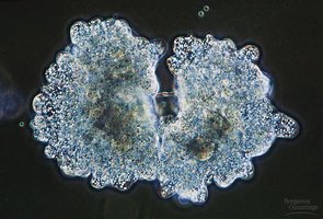
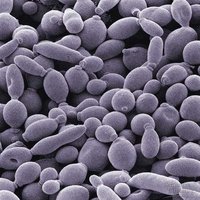
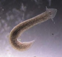
Asexual Reproduction: Involves the creation of genetically identical offspring (clones) without the fusion of gametes. Examples include budding in yeast, fragmentation in lichens, and fission in planaria.
Reasons for Cell Division
Growth and Development: Multicellular organisms grow from a single fertilized egg (zygote) to an adult through repeated cell divisions.
Repair and Maintenance: Damaged or aged cells are replaced by new cells to maintain tissue integrity.
Genetic Material and Chromosomes
During cell division, it is crucial that each daughter cell receives an identical set of genetic information, known as the genome.
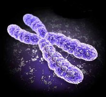
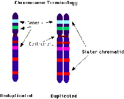
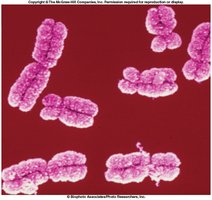
Chromosomes: Structures composed of DNA and proteins (chromatin) that carry genetic information.
Prokaryotes: Typically have one circular chromosome.
Eukaryotes: Have multiple linear chromosomes (e.g., humans have 46 chromosomes, 23 from each parent).
Chromosome Structure
Chromatin: The complex of DNA and proteins that forms chromosomes.
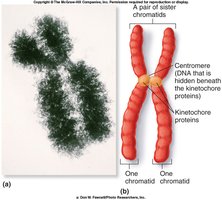
Sister Chromatids: Identical copies of a chromosome joined at a region called the centromere.
The Cell Cycle
Overview of the Cell Cycle
The cell cycle is a series of events that cells go through as they grow and divide. It consists of interphase (cell growth and DNA replication) and the mitotic phase (mitosis and cytokinesis).
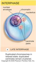
Interphase: The longest phase, where the cell grows, replicates its DNA, and prepares for division.
Mitosis: Division of the nucleus to produce two identical nuclei.
Cytokinesis: Division of the cytoplasm, resulting in two separate cells.
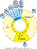
Phases of Interphase
G1 Phase: Cell grows and carries out normal functions.
S Phase: DNA is replicated.
G2 Phase: Further growth and preparation for mitosis.
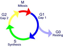
G0 Phase
Some specialized cells exit the cell cycle and enter a resting state called G0. Examples include nerve, adipose, and muscle cells.
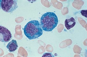
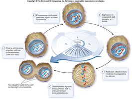
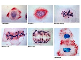
Mitosis
Phases of Mitosis
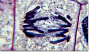
Mitosis is divided into four main phases: prophase, metaphase, anaphase, and telophase (PMAT).
Prophase: Chromosomes condense, nuclear envelope breaks down, spindle forms, centrioles move to poles (plants lack centrioles).
Metaphase: Chromosomes align at the metaphase plate, spindle attaches to centromeres.
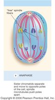
Anaphase: Sister chromatids separate and move to opposite poles.
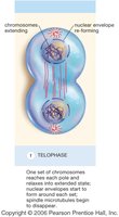
Telophase: Chromosomes decondense, nuclear envelope reforms, cytokinesis begins.
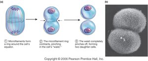
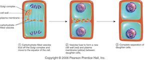
Cytokinesis
Animal Cells: Cleavage furrow forms, pinching the cell into two.
Plant Cells: Cell plate forms, dividing the cell into two daughter cells.
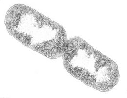
Prokaryotic Cell Division: Binary Fission
Prokaryotes divide by binary fission, a simpler process than mitosis.
One circular chromosome replicates.
Origins move to opposite ends, cell splits into two identical cells.
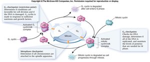
Control of the Cell Cycle
Cell Cycle Checkpoints
Checkpoints regulate the cell cycle, ensuring proper division and preventing errors.
G1/S Checkpoint: Checks cell size, nutrients, and DNA integrity.
G2/M Checkpoint: Ensures DNA is correctly replicated and repaired.
Metaphase/Anaphase Checkpoint: Ensures chromosomes are properly aligned before separation.
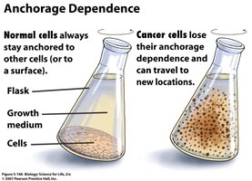
Density-Dependent Inhibition and Anchorage Dependence
Density-Dependent Inhibition: Cells stop dividing when crowded.
Anchorage Dependence: Cells must be attached to a surface to divide.
Contact Inhibition: Cells stop dividing when they touch each other.
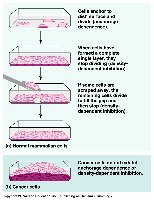
Cancer and Cell Division
Cancer: Loss of Cell Cycle Control
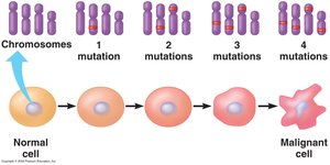
Cancer results from uncontrolled cell division due to mutations that allow cells to bypass checkpoints.
Carcinogens: Substances that cause mutations (e.g., UV light, chemicals, viruses).
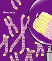
Telomeres: Protective DNA sequences at chromosome ends; shorten with each division. Cancer cells often produce telomerase to maintain telomere length.
Hayflick Limit: Normal human cells divide about 50 times before stopping.
Multi-hit Hypothesis: Multiple mutations are usually required for cancer to develop.

Types of Tumors
Benign Tumor: Non-invasive, does not spread.
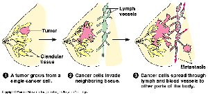
Malignant Tumor: Invades tissues, can spread (metastasize) via lymphatic system.
Metastasis: Spread of cancer cells to distant organs.
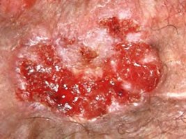
Sunlight and Skin Cancer
UV Light: Causes DNA mutations, immunosuppression, and increases cancer risk.
Types of Skin Cancer:
Basal Cell Carcinoma: Most common, least malignant.
Squamous Cell Carcinoma: Grows rapidly, may metastasize.
Malignant Melanoma: Most dangerous, spreads via lymph system.
ABCDE Rule: Asymmetry, Border irregularity, Color variation, Diameter >6mm, Evolution of lesion.


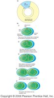
Meiosis: The Formation of Sex Cells
Overview of Meiosis
Meiosis is a specialized form of cell division that produces four genetically unique haploid cells (gametes) from a diploid parent cell. It is essential for sexual reproduction and genetic diversity.
Occurs in: Sex organs (ovaries and testes).
Produces: Sperm in males, eggs in females.
Two Rounds of Division: Meiosis I and Meiosis II, but only one round of DNA replication.
Genetic Variation in Meiosis
Crossing Over: Exchange of genetic material between homologous chromosomes during prophase I, increasing genetic diversity.
Random Alignment: Homologous chromosomes align randomly at metaphase I, leading to many possible combinations.
Phases of Meiosis
Meiosis I: Homologous chromosomes separate, producing two haploid cells.
Meiosis II: Sister chromatids separate, resulting in four unique haploid cells.
Mitosis vs. Meiosis
Feature | Mitosis | Meiosis |
|---|---|---|
Number of Divisions | 1 | 2 |
Number of Daughter Cells | 2 | 4 |
Genetic Identity | Identical | Unique |
Chromosome Number | Diploid (2n) | Haploid (n) |
Function | Growth, repair | Gamete production |
Gametogenesis
Spermatogenesis
Spermatogenesis is the process of sperm formation in males, resulting in four functional, haploid sperm cells from each primary spermatocyte.
1 primary spermatocyte (2n) → 2 secondary spermatocytes (n) → 4 spermatids (n) → 4 spermatozoa
Oogenesis
Oogenesis is the process of egg formation in females, producing one functional ovum and three polar bodies (which degenerate) from each primary oocyte.
Primary oocyte (2n) → 1 secondary oocyte (n) + 1 polar body (n)
Secondary oocyte (n) → 1 ootid (n) + 1 polar body (n)
First polar body may divide to form two more polar bodies
Primary oocytes are arrested in meiosis I at birth; secondary oocytes are arrested in metaphase II until fertilization
Comparison: Spermatogenesis vs. Oogenesis
Feature | Spermatogenesis | Oogenesis |
|---|---|---|
Number of functional gametes | 4 sperm | 1 ovum |
Timing | Continuous after puberty | Begins before birth, completes after fertilization |
Polar bodies | None | 3 (degenerate) |
Sexual Reproduction and Life Cycles
Sexual Reproduction
Sexual reproduction involves the fusion of two haploid gametes from different individuals, resulting in offspring with genetic variation. This variation arises from crossing over, random alignment, and the union of unique gametes.
