 Back
Ch. 7 - Cell Membrane Structure and Function: Study Notes
Study Guide - Smart Notes
 Tailored notes based on your materials, expanded with key definitions, examples, and context.
Cell Membrane Structure & Function
The Fluid Mosaic Model
The fluid mosaic model describes the dynamic and heterogeneous nature of the cell membrane. This model highlights that membranes are not static; instead, they are flexible structures composed of a phospholipid bilayer with embedded proteins, carbohydrates, and cholesterol. The components can move laterally within the layer, allowing for membrane fluidity and adaptability.
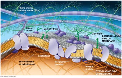
Phospholipid bilayer: Forms the fundamental structure, with hydrophilic heads facing outward and hydrophobic tails inward.
Proteins: Integral and peripheral proteins serve various functions such as transport, signaling, and structural support.
Carbohydrates: Attached to proteins and lipids on the extracellular surface, important for cell recognition.
Cholesterol: Modulates membrane fluidity and stability.
Example: The cell membrane's flexibility allows immune cells to change shape as they move through tissues.
Phospholipid Structure and Membrane Fluidity
Phospholipids are amphipathic molecules with a hydrophilic (water-attracting) head and two hydrophobic (water-repelling) tails. The arrangement of these molecules forms a bilayer, creating a semi-permeable barrier between the cell and its environment.
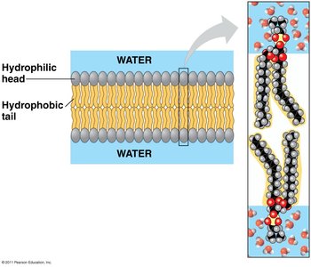
Hydrocarbon chain saturation: Saturated fatty acids make the membrane less fluid, while unsaturated fatty acids (with double bonds) increase fluidity.
Adaptation: Organisms can adjust the saturation of membrane lipids to maintain optimal fluidity under different temperatures.
Example: Fish living in cold water have more unsaturated fatty acids in their membranes to prevent rigidity.
Membrane Proteins and Their Functions
Types and Roles of Membrane Proteins
Membrane proteins are essential for the diverse functions of the cell membrane. They can be classified based on their position and function:
Transport proteins: Facilitate the movement of substances across the membrane (e.g., lac permease for sugar uptake).
Enzymatic proteins: Catalyze reactions at the membrane surface (e.g., adenylate cyclase produces cAMP from ATP).
Signal transduction proteins: Transmit signals from the external environment to the cell's interior (e.g., estrogen receptor).
Cell-cell recognition proteins: Allow cells to identify each other (e.g., CD4 receptor on white blood cells).
Intercellular joining proteins: Connect adjacent cells (e.g., connexin in heart muscle).
Attachment proteins: Anchor the membrane to the cytoskeleton and extracellular matrix (e.g., integrin).
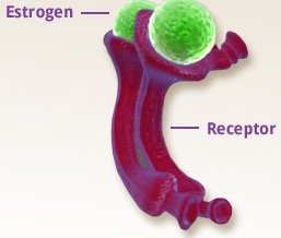
Example: The estrogen receptor is a membrane protein that binds estrogen, triggering changes in gene expression.
Membrane Sidedness and Assembly
How Membrane Sidedness Arises
Membrane sidedness refers to the distinct composition and orientation of the inner and outer leaflets of the lipid bilayer. This asymmetry is established during membrane synthesis in the endoplasmic reticulum and Golgi apparatus and is maintained as vesicles fuse with the plasma membrane.
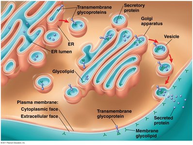
Glycoproteins and glycolipids: Carbohydrate groups are added in the ER and Golgi, facing the extracellular side after vesicle fusion.
Functional significance: Sidedness is crucial for processes like cell signaling and recognition.
Transport Across Cell Membranes
Passive and Active Transport Mechanisms
Transport across the cell membrane is essential for maintaining homeostasis. There are two main categories:
Passive transport: Movement of substances down their concentration gradient without energy input. Includes simple diffusion and facilitated diffusion.
Active transport: Movement of substances against their concentration gradient, requiring energy (usually ATP) and specific transport proteins.

Example: The sodium-potassium pump is an active transporter that maintains ion gradients in animal cells.
Simple Diffusion and Facilitated Diffusion
Simple diffusion is the movement of small, nonpolar molecules (e.g., O2, CO2) directly through the lipid bilayer. Facilitated diffusion involves transport proteins that help polar or charged molecules cross the membrane.
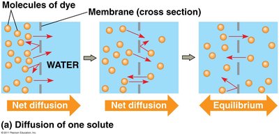
Equilibrium: Net movement continues until concentrations are equal on both sides.
Transport proteins: Channel and carrier proteins enable facilitated diffusion.
Osmosis and Tonicity
Osmosis: Water Movement Across Membranes
Osmosis is the diffusion of water across a selectively permeable membrane in response to differences in solute concentration or pressure. Water moves through specialized channels called aquaporins.
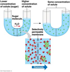
Direction of water movement: Water moves from areas of lower solute concentration to higher solute concentration.
Biological importance: Osmosis regulates cell volume and internal environment.
Tonicity: Effects of Solute Concentration on Cells
Tonicity describes how a solution affects cell volume due to water movement. It depends on the concentration of nonpenetrating solutes and membrane permeability.
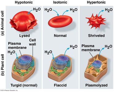
Hypotonic solution: Lower solute concentration outside; water enters the cell, causing swelling or lysis.
Isotonic solution: Equal solute concentration; no net water movement, cell remains normal.
Hypertonic solution: Higher solute concentration outside; water leaves the cell, causing shrinkage or plasmolysis.
Example: Red blood cells placed in pure water (hypotonic) will swell and burst.
Ion Channels and Pain Perception
Role of Ion Channels in Sensory Function
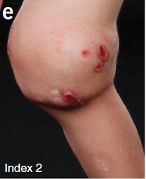
Ion channels are membrane proteins that allow specific ions to pass through the membrane, playing a critical role in nerve impulse transmission and sensory perception. Mutations in these channels can lead to altered pain perception or disease.
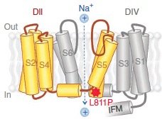
Example: A mutation in a sodium channel gene can result in congenital insensitivity to pain, leading to repeated injuries.
Active Transport and Cotransport
Active Transport Mechanisms
Active transport uses energy (often from ATP) to move substances against their concentration gradients. This process is essential for maintaining cellular ion balances and nutrient uptake.
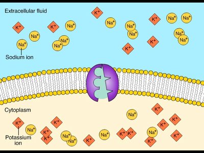
Example: The sodium-potassium pump exchanges Na+ and K+ ions across the plasma membrane.
Cotransport: Coupled Movement of Substances
Cotransport involves the simultaneous movement of two substances across the membrane, often coupling the downhill movement of one molecule with the uphill movement of another.
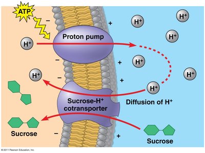
Symport: Both substances move in the same direction.
Antiport: Substances move in opposite directions.
Example: The sucrose-H+ cotransporter in plants uses the proton gradient to import sucrose into cells.
Bulk Transport Across the Plasma Membrane
Endocytosis and Exocytosis
Bulk transport mechanisms move large particles or volumes of substances across the membrane. Endocytosis brings materials into the cell via vesicles, while exocytosis expels materials out of the cell.
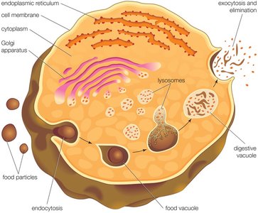
Phagocytosis: Cell engulfs large particles or cells ("cell eating").
Pinocytosis: Cell takes in extracellular fluid ("cell drinking").
Receptor-mediated endocytosis: Specific molecules are taken in after binding to receptors.
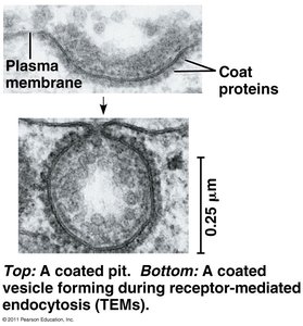
Example: White blood cells use phagocytosis to engulf and destroy pathogens.
