 Back
Cell Membranes: Structure, Function, and Transport Mechanisms
Study Guide - Smart Notes
 Tailored notes based on your materials, expanded with key definitions, examples, and context.
Cell Membranes
Introduction to Cell Membranes
The cell membrane, also known as the plasma membrane, is a fundamental structure that defines the boundary of the cell and regulates the movement of substances into and out of the cell. Its unique composition and properties are essential for maintaining cellular homeostasis and enabling communication with the environment.
Constraints on Cell Size
Surface Area to Volume Ratio
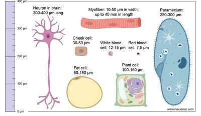
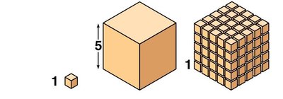
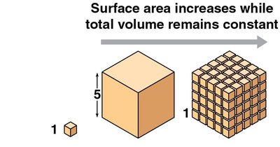
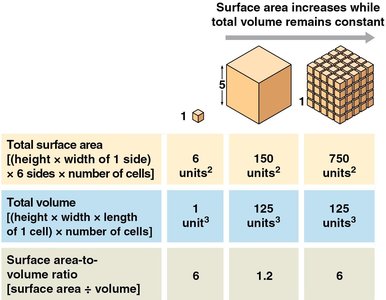
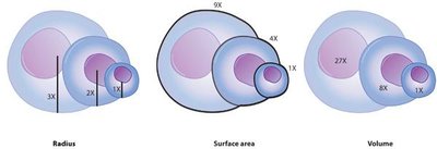
Cell size is limited by the relationship between surface area and volume. As a cell grows, its volume increases faster than its surface area, which restricts the rate at which materials can enter or leave the cell. This constraint is a key reason why most cells are microscopic.
Surface Area: The total area available for exchange of materials.
Volume: The total space inside the cell, representing the demand for resources and waste removal.
Surface Area to Volume Ratio: As cell size increases, this ratio decreases, limiting efficient transport.
Example: Many small cells have a greater total surface area than one large cell of the same volume, facilitating more efficient exchange of materials.
The Structure of Cell Membranes
The Fluid Mosaic Model
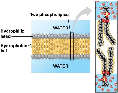
The fluid mosaic model describes the structure of the plasma membrane as a dynamic arrangement of phospholipids, proteins, and carbohydrates. Phospholipids form a bilayer with hydrophilic heads facing outward and hydrophobic tails inward, creating a semi-permeable barrier.
Phospholipids: Amphipathic molecules with hydrophilic heads and hydrophobic tails.
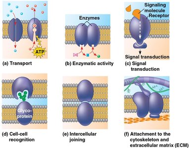
Membrane Proteins: Embedded or associated proteins that perform various functions such as transport, signaling, and structural support.
Carbohydrates: Attached to lipids or proteins, contributing to cell recognition and signaling.

Membrane Fluidity
Factors Influencing Membrane Fluidity
Membrane fluidity is crucial for proper membrane function and is influenced by several factors:
Hydrocarbon Tail Structure: Unsaturated fatty acids increase fluidity, while saturated fatty acids decrease it.
Cholesterol Content: Cholesterol acts as a fluidity buffer, preventing membranes from becoming too rigid or too fluid.
Temperature: Higher temperatures increase fluidity; lower temperatures decrease it.
Species Adaptations: Organisms living in extreme environments have unique membrane compositions to maintain optimal fluidity.
Transport Across the Cell Membrane
Overview of Transport Mechanisms
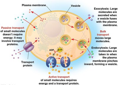
The plasma membrane regulates the movement of substances through several mechanisms, classified as passive transport, active transport, and bulk transport.
Passive Transport: Movement of molecules down their concentration gradient without energy input.
Active Transport: Movement of molecules against their concentration gradient, requiring energy (usually ATP).
Bulk Transport: Movement of large particles or volumes via vesicles (endocytosis and exocytosis).
Passive Transport
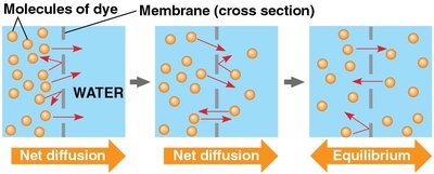
Passive transport includes simple diffusion, facilitated diffusion, and osmosis.
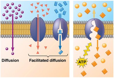
Simple Diffusion: Movement of small, nonpolar molecules directly through the lipid bilayer.
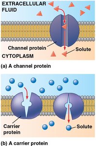
Facilitated Diffusion: Movement of polar or charged molecules via channel or carrier proteins.
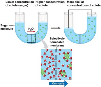
Osmosis: Diffusion of water across a selectively permeable membrane.
Facilitated Diffusion
Facilitated diffusion uses membrane proteins to transport substances that cannot diffuse directly through the lipid bilayer.
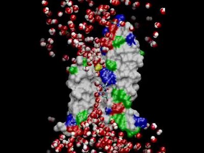
Channel Proteins: Provide hydrophilic tunnels for specific molecules or ions (e.g., aquaporins for water).
Carrier Proteins: Bind to molecules and change shape to shuttle them across the membrane.
Osmosis
Osmosis is the diffusion of water across a selectively permeable membrane. Water moves from areas of lower solute concentration to higher solute concentration to balance solute levels.
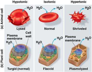
Hypotonic Solution: Lower solute concentration outside; water enters the cell.
Isotonic Solution: Equal solute concentration; no net water movement.
Hypertonic Solution: Higher solute concentration outside; water leaves the cell.
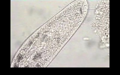
Example: Paramecium uses a contractile vacuole to expel excess water in hypotonic environments.
Active Transport
Active transport moves substances against their concentration gradients, requiring energy input, usually from ATP. This process allows cells to maintain internal concentrations of small molecules that differ from those in the environment.
Carrier Proteins: Specialized proteins that use energy to transport substances (e.g., sodium-potassium pump).
Example: The sodium-potassium (Na-K) pump moves Na+ out and K+ into animal cells, maintaining membrane potential.
Membrane Potential: The voltage difference across a membrane, created by ion gradients.
Electrochemical Gradient: Combination of concentration gradient and electrical charge difference.
Bulk Transport
Bulk transport moves large molecules or particles via vesicles. This includes endocytosis (import) and exocytosis (export).
Endocytosis: The cell engulfs material, forming a vesicle that is brought into the cell.
Exocytosis: Vesicles fuse with the plasma membrane to release contents outside the cell.
Phagocytosis: "Cell eating"—engulfing large particles.
Pinocytosis: "Cell drinking"—engulfing extracellular fluid.
Summary Table: Types of Membrane Transport
Type | Energy Required? | Direction | Example |
|---|---|---|---|
Simple Diffusion | No | Down gradient | O2, CO2 |
Facilitated Diffusion | No | Down gradient | Glucose via carrier protein |
Osmosis | No | Down water potential | Water via aquaporin |
Active Transport | Yes (ATP) | Against gradient | Na-K pump |
Bulk Transport | Yes (ATP) | In or out | Endocytosis, exocytosis |
Key Equations
Surface Area of a Cube:
Volume of a Cube:
Surface Area to Volume Ratio:
Conclusion
The cell membrane is a dynamic and complex structure essential for maintaining cellular integrity, regulating transport, and facilitating communication. Understanding its properties and mechanisms is fundamental to cell biology and physiology.
