 Back
BackCell Specialization and Animal Development
Study Guide - Smart Notes
 Tailored notes based on your materials, expanded with key definitions, examples, and context.
Cell Specialization and Animal Development
Overview of Cell Specialization
Cell specialization is a fundamental process in multicellular organisms, allowing genetically identical cells to develop distinct structures and functions. This process is orchestrated by differential gene expression during embryonic development, leading to the formation of tissues, organs, and organ systems.
Cell specialization enables the formation of complex organisms from a single fertilized egg (zygote).
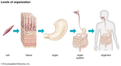
Cells are organized hierarchically: cells → tissues → organs → organ systems → organism.
Gene Expression and Cell Specialization
Gene expression is the process by which information from a gene is used to synthesize functional gene products (proteins or RNAs), which determine cell structure and function. During development, gene expression is tightly regulated, resulting in cell differentiation and morphogenesis.
Cell division (mitosis) produces genetically identical cells.
Cell differentiation is the process by which unspecialized cells become specialized in structure and function.
Morphogenesis refers to the development of the organism's shape and structure.
Regulation of Gene Expression: Cytoplasmic Determinants and Induction
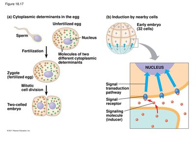
Two major factors influence gene expression during early development:
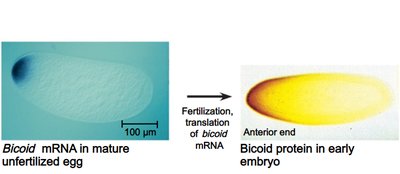
Cytoplasmic determinants: Maternal substances in the egg that are unevenly distributed and influence the fate of cells after fertilization.
Induction: The process by which signaling molecules from one group of embryonic cells influence the gene expression of neighboring cells, leading to cell differentiation.
Determination and Differentiation
Cell specialization proceeds through stages:
Determination: The process by which a cell becomes committed to a specific fate, often before it shows any visible signs of differentiation.
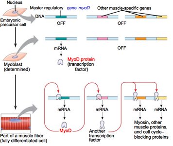
Differentiation: The process by which a determined cell develops into its final specialized form, marked by the production of tissue-specific proteins.
Example: Muscle cell differentiation involves the activation of the myoD gene, which encodes a transcription factor that initiates the muscle-specific gene expression program.
Stem Cells and Potency
Types of Stem Cells
Stem cells are undifferentiated cells with the capacity to divide and give rise to specialized cell types. Their potential is classified by potency:
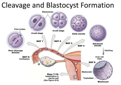
Totipotent: Can give rise to all cell types of the organism, including extraembryonic tissues (e.g., fertilized egg, early morula).
Pluripotent: Can differentiate into all body cell types but not extraembryonic tissues (e.g., inner cell mass of the blastocyst).
Multipotent: Can develop into a limited range of cell types within a particular tissue or organ (e.g., hematopoietic stem cells in bone marrow).
Induced pluripotent stem cells (iPSCs) are adult cells reprogrammed to a pluripotent state, a discovery awarded the Nobel Prize in 2012.

Animal Development: Early Embryogenesis
Stages of Early Embryonic Development
Animal development follows a conserved sequence of stages:
Fertilization: Fusion of egg and sperm to form a zygote.
Cleavage: Rapid mitotic divisions partition the zygote into smaller cells without increasing overall size, forming the morula (solid ball of cells).
Blastulation: Formation of the blastula, a hollow sphere of cells with an inner cell mass (embryoblast) and an outer trophoblast. The blastocoel is the fluid-filled cavity.
Gastrulation: Rearrangement of the blastula into a three-layered structure (ectoderm, mesoderm, endoderm) through cell migration and invagination, forming the archenteron (primitive gut) and blastopore (future anus).
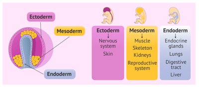
The Three Embryonic Germ Layers
Gastrulation produces three primary germ layers, each giving rise to specific tissues and organs:
Ectoderm (outer layer): Forms the epidermis, nervous system, and related structures.
Mesoderm (middle layer): Forms muscles, bones, connective tissue, circulatory system, and reproductive organs.
Endoderm (inner layer): Forms the lining of the digestive tract, lungs, and associated organs.
Morphogenesis and Pattern Formation
Pattern Formation and Positional Information
Pattern formation is the spatial organization of tissues and organs during development. Cells receive positional information—molecular cues that inform them of their location relative to body axes and neighboring cells, guiding their differentiation and organization.
Genetic Control of Pattern Formation: Drosophila as a Model
Research in Drosophila melanogaster (fruit fly) has revealed key genes controlling pattern formation:
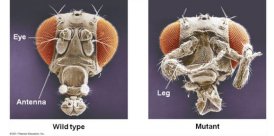
Homeotic genes: Control the identity of body segments; mutations can cause body parts to develop in the wrong location (e.g., legs instead of antennae).
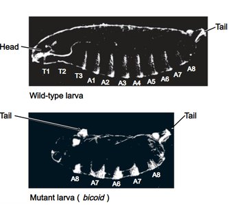
Maternal effect genes (egg-polarity genes): Such as the bicoid gene, establish the anterior-posterior axis of the embryo. Loss of bicoid results in embryos lacking anterior structures.
Summary Table: Types of Stem Cells and Their Potency
Stem Cell Type | Potency | Examples |
|---|---|---|
Totipotent | All cell types, including extraembryonic tissues | Zygote, early morula |
Pluripotent | All body cell types | Inner cell mass of blastocyst, iPSCs |
Multipotent | Multiple cell types within a tissue | Hematopoietic stem cells |
Unipotent | One cell type | Muscle stem cells |
Key Equations and Concepts
Gene expression regulation:
Cleavage:
