 Back
BackCell Structure and Function: A Comprehensive Study Guide
Study Guide - Smart Notes
 Tailored notes based on your materials, expanded with key definitions, examples, and context.
Cell Structure and Function
4.1 Cellular Level of Organization
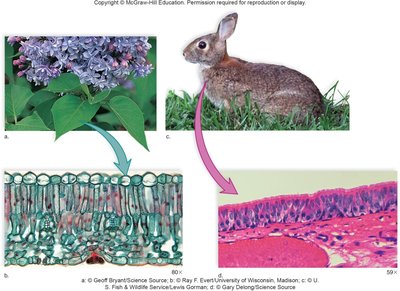
The cell is the fundamental unit of life, forming the basis for all living organisms. The development of cell theory in the 19th century unified biological sciences and established that all organisms are composed of cells, all cells arise from preexisting cells, and cells are the basic units of structure and function in organisms.
Cell Theory: Formulated by Schleiden, Schwann, and Virchow, it states:
All living things are composed of cells.
Cells arise only from preexisting cells.
Cells are the basic units of structure and function in organisms.
Historical Context: The cell theory was developed through the work of botanists and zoologists in the 1830s.
Cell Size and Surface Area-to-Volume Ratio
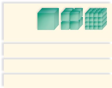
Cells vary in size but are generally small to maximize their surface area-to-volume ratio, which is critical for efficient exchange of materials with the environment.
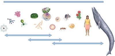
Cell Size: Ranges from 1 mm to 1 μm in diameter.
Surface Area-to-Volume Ratio: As cells increase in size, their volume grows faster than their surface area, reducing efficiency in material exchange.
Importance: Small cells have a larger surface area relative to volume, facilitating efficient transport of molecules.
Microscopy in Cell Biology
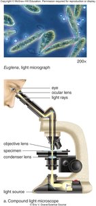
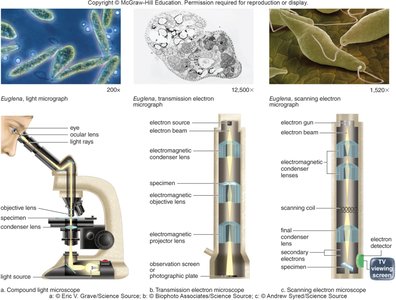
Microscopy is essential for studying cells, as most are too small to be seen with the naked eye. Different types of microscopes provide varying levels of magnification and resolution.
Compound Light Microscope: Uses light and glass lenses to magnify specimens up to 1000X; resolves objects separated by 0.2 μm.
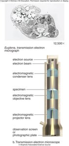
Transmission Electron Microscope (TEM): Uses electrons and magnetic lenses for much higher magnification and resolution (up to 100,000X; resolves objects separated by 0.0002 μm).
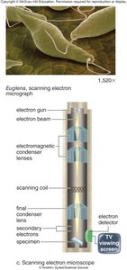
Scanning Electron Microscope (SEM): Provides 3D images of specimen surfaces by detecting emitted electrons.
Key Terms: Magnification (image size vs. actual size), Resolution (minimum distance to distinguish two points), Contrast (difference in shading).
The Plasma Membrane
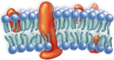
The plasma membrane is a universal feature of all cells, forming a selective barrier that regulates the movement of substances in and out of the cell.
Structure: Composed of a phospholipid bilayer with embedded proteins.
Function: Maintains homeostasis by controlling the internal environment of the cell.
4.2 Prokaryotic Cells
Characteristics and Classification
Prokaryotic cells lack a membrane-bound nucleus and are structurally simpler than eukaryotic cells. They are classified into two domains: Bacteria and Archaea.
Bacteria: Can cause disease, decompose organic material, and are used in biotechnology.
Archaea: Often inhabit extreme environments and differ biochemically from bacteria.
Prokaryotic Cell Structure
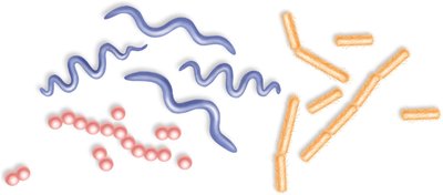
Shapes: Spherical (coccus), rod-shaped (bacillus), spiral (spirillum or spirochete).
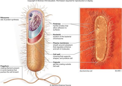
Cell Envelope: Includes plasma membrane, cell wall (with peptidoglycan), and sometimes a capsule (polysaccharide layer).
Cytoplasm: Contains water, enzymes, and molecules; nucleoid region holds circular DNA; plasmids are small DNA rings; ribosomes synthesize proteins.
External Structures: Flagella (motility), fimbriae (attachment), conjugation pili (DNA transfer).
4.3 Introduction to Eukaryotic Cells
General Features
Eukaryotic cells are characterized by a membrane-bound nucleus, specialized organelles, and a complex internal structure. They are generally larger and more compartmentalized than prokaryotic cells.
Organelles: Specialized structures performing specific functions, often isolated by membranes.
Plasma Membrane: Separates cell from environment and regulates material passage.
Compartmentalization: Allows for specialization and efficiency in cellular processes.
Origin of Eukaryotic Organelles
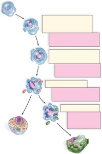
The endosymbiotic theory explains the origin of mitochondria and chloroplasts as formerly free-living prokaryotes engulfed by ancestral eukaryotic cells.
Nucleus: Likely evolved from plasma membrane invagination.
Endomembrane System: Developed from membrane proliferation.
Mitochondria and Chloroplasts: Resulted from endosymbiosis, providing metabolic advantages.
Animal and Plant Cell Anatomy
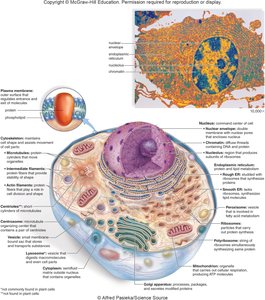
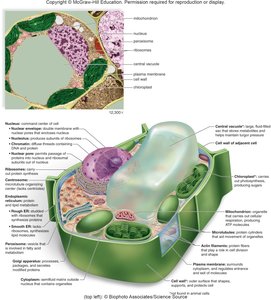
Animal and plant cells share many organelles but also have unique features. Plant cells possess cell walls and chloroplasts, while animal cells have centrioles and lysosomes.
4.4 The Nucleus and Ribosomes
The Nucleus
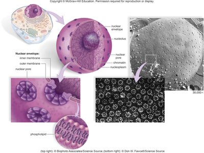
The nucleus is the command center of the cell, housing genetic material and coordinating cellular activities.
Nuclear Envelope: Double membrane with pores for exchange between nucleoplasm and cytoplasm.
Chromatin: DNA-protein complex that condenses into chromosomes during cell division.
Nucleolus: Site of ribosomal RNA (rRNA) synthesis and ribosome subunit assembly.
Ribosomes
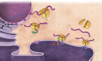
Ribosomes are the sites of protein synthesis, composed of rRNA and proteins. They may be free in the cytoplasm or bound to the endoplasmic reticulum (ER).
Structure: Consist of large and small subunits assembled in the nucleolus.
Function: Translate mRNA into polypeptides; free ribosomes synthesize cytoplasmic proteins, while ER-bound ribosomes synthesize proteins for membranes or export.
Central Dogma: Information flows from DNA to mRNA to protein.
4.5 The Endomembrane System
Components and Functions
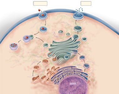
The endomembrane system is a network of membranes within the cell that compartmentalizes functions and facilitates transport of molecules.
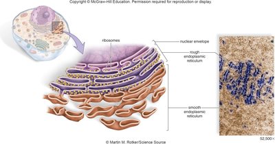
Includes: Nuclear envelope, endoplasmic reticulum (ER), Golgi apparatus, and vesicles.
Function: Restricts enzymatic reactions to specific compartments, sorts and ships proteins and lipids.
Endoplasmic Reticulum (ER)
Rough ER: Studded with ribosomes; synthesizes and processes proteins, forms glycoproteins, and produces transport vesicles.
Smooth ER: Lacks ribosomes; synthesizes lipids, detoxifies chemicals, and stores substances.
Golgi Apparatus
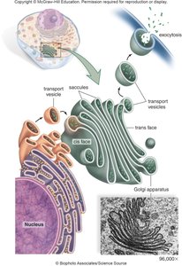
The Golgi apparatus modifies, sorts, and packages proteins and lipids for transport within or outside the cell.
Structure: Stack of flattened, curved saccules.
Function: Receives vesicles from ER, modifies contents, and directs them to their destinations.
Lysosomes
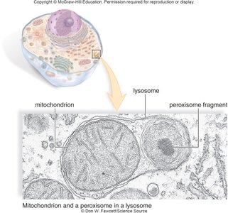
Lysosomes are membrane-bound vesicles containing digestive enzymes, responsible for breaking down macromolecules and recycling cellular components.
Function: Digest large molecules, recycle resources, and destroy pathogens in white blood cells.
Clinical Relevance: Lysosomal storage diseases (e.g., Tay-Sachs) result from enzyme defects.
Endomembrane System Summary
Proteins and lipids produced in the ER are modified and sorted by the Golgi apparatus, then transported in vesicles to their final destinations, including secretion or lysosomal digestion.
4.6 Microbodies and Vacuoles
Microbodies (Peroxisomes)
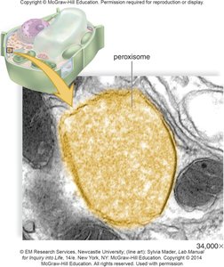
Microbodies are specialized vesicles containing enzymes for specific metabolic functions, such as breaking down fatty acids and detoxifying hydrogen peroxide.
Peroxisomes: Contain enzymes that produce and degrade hydrogen peroxide; active in lipid metabolism.
Clinical Note: Defects in peroxisomal proteins can cause neurological disorders (e.g., ALD).
Vacuoles
Vacuoles are large membranous sacs for storage and maintenance of cell turgor. Plant cells have a prominent central vacuole that stores water, nutrients, and waste products.
Functions: Storage, development of turgor pressure, and breakdown of aged organelles.
4.7 Energy-Related Organelles
Chloroplasts
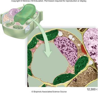
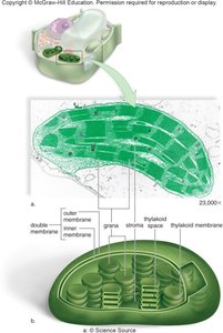
Chloroplasts are the sites of photosynthesis in plants and algae, converting solar energy into chemical energy stored in carbohydrates.
Structure: Double membrane, internal thylakoid membranes stacked into grana, and stroma (fluid matrix).
Function: Photosynthesis:
Chlorophyll: Green pigment located in thylakoid membranes.
Mitochondria
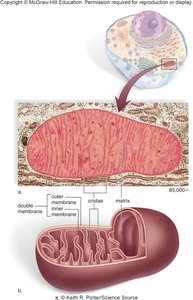
Mitochondria are the powerhouses of the cell, generating ATP through cellular respiration. They are present in nearly all eukaryotic cells.
Structure: Double membrane; inner membrane forms cristae, enclosing the matrix with respiratory enzymes.
Function: Break down carbohydrates, use oxygen, and produce ATP.
Endosymbiotic Theory: Mitochondria contain their own DNA and ribosomes, supporting their evolutionary origin as free-living bacteria.
4.8 The Cytoskeleton
Overview
The cytoskeleton is a dynamic network of protein fibers that maintains cell shape, enables movement, and organizes cell contents.
Types of Fibers: Actin filaments, intermediate filaments, microtubules.
Functions: Structural support, intracellular transport, cell division, and motility.
Actin Filaments
Structure: Thin, twisted filaments beneath the plasma membrane.
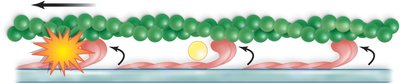
Functions: Maintain cell shape, support microvilli, enable cytoplasmic streaming, and muscle contraction (with myosin).
Intermediate Filaments
Structure: Rope-like fibers of varying composition.
Functions: Support nuclear envelope, reinforce cell junctions, and provide mechanical strength (e.g., keratin in skin).
Microtubules
Structure: Hollow cylinders of α- and β-tubulin dimers, organized by the centrosome (MTOC).
Functions: Organelle movement (with kinesin and dynein), mitotic spindle formation during cell division.
Centrioles
Structure: Short cylinders of microtubules arranged in 9 triplets; found in animal cells.
Functions: Organize microtubules during cell division, give rise to basal bodies of cilia and flagella.
Cilia and Flagella
Cilia and flagella are hair-like projections that facilitate cell movement. They have a characteristic "9+2" arrangement of microtubules.
Cilia: Short, numerous, move in coordinated waves (e.g., respiratory tract).
Flagella: Longer, usually singular, move in a whip-like or propeller motion (e.g., sperm cells).
Additional info: This guide expands on the lecture outline with academic context, definitions, and examples to provide a comprehensive, self-contained resource for college-level biology students.
