 Back
BackCell Structure and Function: A Comprehensive Overview
Study Guide - Smart Notes
 Tailored notes based on your materials, expanded with key definitions, examples, and context.
Cell Structure and Function
Overview of Cell Biology
Cells are the fundamental units of life, forming the basis of structure and function in all living organisms. This chapter explores the tools used to study cells, the differences between prokaryotic and eukaryotic cells, and the organization and function of cellular components.
Microscopy and Cell Study
Microscopy: Visualizing Cells
Microscopes are essential for studying cells, which are typically too small to be seen by the naked eye. The three main parameters of microscopy are:
Magnification: The ratio of an object's image size to its real size.
Resolution: The clarity of the image, or the minimum distance between two distinguishable points.
Contrast: Visible differences in brightness between parts of the sample.
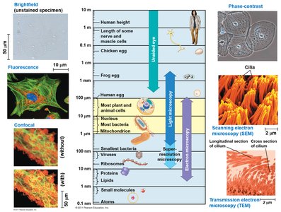
Types of Microscopy: Light microscopy, fluorescence, confocal, phase-contrast, scanning electron microscopy (SEM), and transmission electron microscopy (TEM) are used to visualize different aspects of cell structure and function.
Cell Fractionation
Cell fractionation is a technique used to separate cellular components for detailed study. This process involves homogenization and centrifugation, allowing scientists to isolate organelles and analyze their functions.
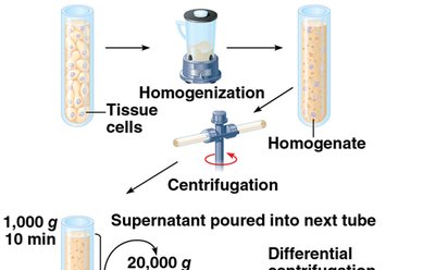
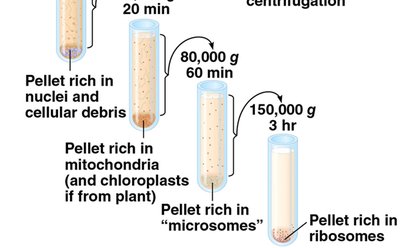
Homogenization: Breaking open cells to release their contents.
Centrifugation: Spinning the homogenate at various speeds to separate components by size and density.
Cell Types and Basic Features
Prokaryotic vs. Eukaryotic Cells
All cells share certain features, but are classified as prokaryotic or eukaryotic based on structural differences.
Prokaryotic cells: Lack a nucleus and membrane-bound organelles. DNA is located in the nucleoid region. Examples include Bacteria and Archaea.
Eukaryotic cells: Have a nucleus enclosed by a nuclear envelope and possess membrane-bound organelles. Examples include protists, fungi, animals, and plants.
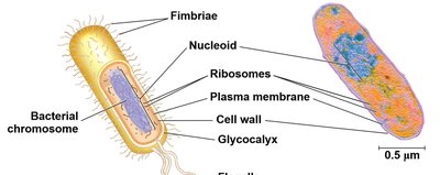

Basic Features of All Cells
Plasma membrane: Selective barrier regulating passage of substances.
Cytosol: Semifluid substance within the cell.
Chromosomes: Carry genetic information.
Ribosomes: Synthesize proteins.
Plasma Membrane Structure
Composition and Function
The plasma membrane is a phospholipid bilayer with embedded proteins, functioning as a selective barrier for the cell.
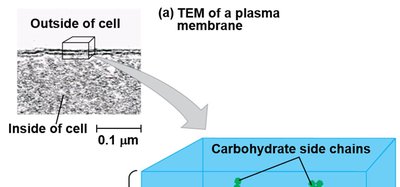
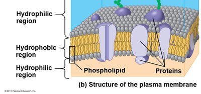
Phospholipid bilayer: Provides fluidity and flexibility.
Proteins: Facilitate transport, signaling, and structural support.
Surface Area to Volume Ratio
A high surface-to-volume ratio is crucial for efficient exchange of materials between the cell and its environment. As cells increase in size, their volume grows faster than their surface area, limiting the rate of exchange.
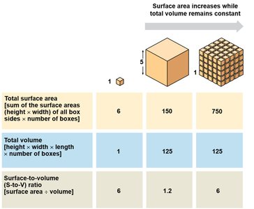
Cube Size | Total Surface Area | Total Volume | Surface-to-Volume Ratio |
|---|---|---|---|
1x1x1 | 6 | 1 | 6 |
5x5x5 | 150 | 125 | 1.2 |
Multiple 1x1x1 | 750 | 125 | 6 |
Internal Organization of Eukaryotic Cells
Animal and Plant Cell Structures
Both animal and plant cells contain most of the same organelles, but some are unique to each type.
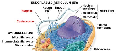
Animal cells: Lysosomes, centrosomes with centrioles, flagella (in some sperm).
Plant cells: Chloroplasts, central vacuole, cell wall, plasmodesmata.
Nucleus and Ribosomes
The Nucleus: Information Center
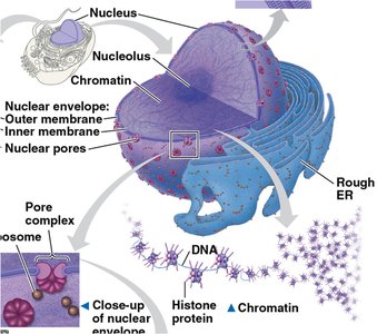
The nucleus contains most of the cell's DNA and is surrounded by a double membrane called the nuclear envelope. Nuclear pores regulate molecular traffic, and the nuclear lamina maintains nuclear shape.
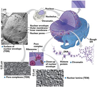

Chromatin: DNA-protein complex that condenses to form chromosomes during cell division.
Nucleolus: Site of ribosomal RNA (rRNA) synthesis.
Ribosomes: Protein Factories
Ribosomes are complexes of rRNA and proteins that synthesize proteins. They can be free in the cytosol or bound to the endoplasmic reticulum (ER) or nuclear envelope.
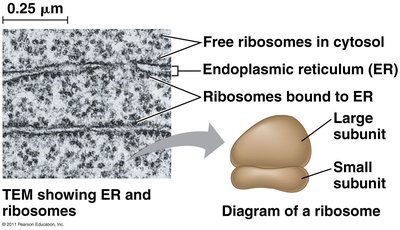
Free ribosomes: Synthesize proteins for use within the cytosol.
Bound ribosomes: Synthesize proteins for membranes, organelles, or export.
The Endomembrane System
Components and Functions
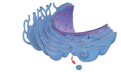
The endomembrane system includes the nuclear envelope, endoplasmic reticulum (ER), Golgi apparatus, lysosomes, vacuoles, and plasma membrane. These structures are either continuous or connected via vesicles, coordinating protein traffic and metabolic functions.
Endoplasmic Reticulum (ER)
The ER is a network of membranes involved in protein and lipid synthesis. It is divided into:
Smooth ER: Synthesizes lipids, detoxifies drugs, stores calcium ions.
Rough ER: Studded with ribosomes, synthesizes glycoproteins, distributes transport vesicles, and produces membranes.
Golgi Apparatus
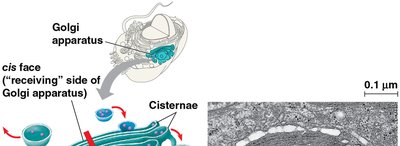
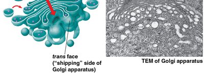
The Golgi apparatus consists of flattened sacs (cisternae) and functions as the cell's shipping and receiving center. It modifies ER products, manufactures macromolecules, and sorts/packages materials into vesicles.
Lysosomes
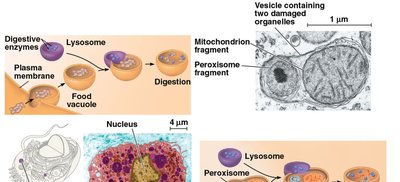
Lysosomes are membranous sacs containing hydrolytic enzymes for digesting macromolecules. They function in phagocytosis (engulfing food particles) and autophagy (recycling cellular components).
Vacuoles
Vacuoles are large vesicles with diverse functions:
Food vacuoles: Formed by phagocytosis.
Contractile vacuoles: Pump excess water out of cells (in protists).
Central vacuole: Stores organic compounds and water (in plants).
Energy Conversion Organelles
Mitochondria and Chloroplasts
Mitochondria are the sites of cellular respiration, generating ATP using oxygen. Chloroplasts, found in plants and algae, are the sites of photosynthesis. Both organelles contain their own DNA and ribosomes, supporting the endosymbiont theory of their origin.
Mitochondria: Have a double membrane, with the inner membrane folded into cristae to increase surface area for ATP synthesis.
Chloroplasts: Contain thylakoids (stacked into grana) and stroma, and are specialized plastids involved in photosynthesis.
Peroxisomes
Peroxisomes are single-membrane organelles that carry out oxidation reactions, producing hydrogen peroxide and converting it to water. Specialized peroxisomes (glyoxysomes) convert fatty acids to sugars in seedlings.
The Cytoskeleton
Structure and Function
The cytoskeleton is a network of protein fibers that organizes cell structure, anchors organelles, and facilitates cell movement. It consists of:
Microtubules: Hollow rods that shape the cell, guide organelle movement, and separate chromosomes during cell division.
Microfilaments (actin filaments): Solid rods that support cell shape and are involved in cell motility and muscle contraction.
Intermediate filaments: Provide mechanical support and anchor organelles.
Centrosomes and Centrioles
Microtubules grow out from centrosomes, which are microtubule-organizing centers. In animal cells, centrosomes contain a pair of centrioles arranged in a ring.
Cilia and Flagella
Microtubules control the movement of cilia and flagella, which are locomotor appendages. They share a common structure: a core of microtubules sheathed by the plasma membrane, anchored by a basal body, and powered by dynein motor proteins.
Extracellular Components and Cell Connections
Cell Walls of Plants
Plant cell walls provide protection, maintain shape, and prevent excessive water uptake. They are composed of cellulose fibers and may have multiple layers (primary wall, middle lamella, secondary wall). Plasmodesmata are channels between plant cells for communication.
Extracellular Matrix (ECM) of Animal Cells
Animal cells lack cell walls but are surrounded by an ECM made of glycoproteins (collagen, proteoglycans, fibronectin). The ECM binds to integrins in the plasma membrane, influencing cell behavior and gene activity.
Intercellular Junctions
Cells in tissues communicate and adhere through specialized junctions:
Plasmodesmata: Channels in plant cell walls for transport and communication.
Tight junctions: Prevent leakage of extracellular fluid in animal cells.
Desmosomes: Anchor cells together into strong sheets.
Gap junctions: Provide cytoplasmic channels between adjacent animal cells.
Integration of Cellular Components
Coordination of Cell Activities
Cells function as integrated units, with organelles and structures working together. For example, a macrophage's ability to destroy bacteria involves coordination between the cytoskeleton, lysosomes, and plasma membrane.
