 Back
Cell Structure and Function: Animal and Plant Cells
Study Guide - Smart Notes
 Tailored notes based on your materials, expanded with key definitions, examples, and context.
Cell Components and Organization
Levels of Organization in Life
Biological identity is determined by the relationship between different levels of organization in the body, from molecules to organelles, cells, tissues, organs, and organisms. These levels, while differing in complexity, share patterns and functions with all life on earth.
Animal and Plant Cell Structure
Cells are the basic units of life. Both animal and plant cells share common features but also have distinct organelles adapted to their functions.
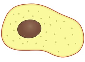
Animal Cell Structure
Nucleus: Control center containing genetic material (DNA).
Cell Membrane: Flexible boundary controlling entry and exit of substances.
Cytoplasm: Jelly-like substance where organelles are suspended and chemical reactions occur.
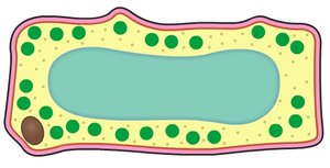
Plant Cell Structure
Nucleus: Contains DNA.
Chloroplast: Site of photosynthesis.
Vacuole: Large central storage sac.
Cytoplasm: Site for chemical reactions.
Cell Wall: Rigid, structural layer outside the membrane.
Cell Membrane: Controls movement of substances.
Comparing Animal and Plant Cells
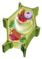
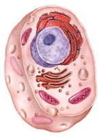
Animal and plant cells differ in several key organelles and functions:
Cell Wall: Present in plant cells, absent in animal cells.
Chloroplasts: Present in plant cells for photosynthesis.
Vacuole: Large and permanent in plant cells, small and temporary in animal cells.
Nucleus and Genetic Material
Nucleus Structure and Function
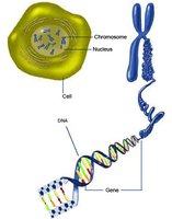
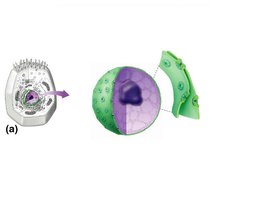
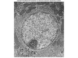
The nucleus is the control center of the cell, containing genetic material (DNA) organized into chromosomes. It is surrounded by a nuclear envelope and contains a dense area called the nucleolus.
Genetic Material: DNA makes up genes on chromosomes, carrying coded information for cell activities and characteristics.
Nuclear Envelope: Double membrane with nuclear pores for transport.
Nucleolus: Site of ribosome synthesis.
Chromosomes and DNA
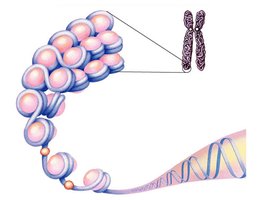
Chromosomes: Visible during cell division, composed of DNA and histone proteins.
DNA: Carries genetic instructions for life.
Cytoplasm
Structure and Function
The cytoplasm is a semi-fluid substance containing water, sugars, salts, and dissolved substances. It is the site for chemical reactions and suspends cell organelles.
Structure: Ranges from liquid to jelly-like.
Function: Site for metabolic reactions; suspends organelles.
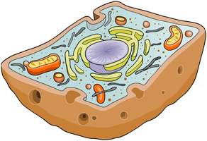
Cell Membrane
Structure and Function
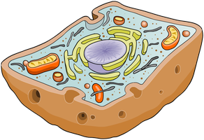
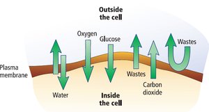
The cell membrane is a flexible, semi-permeable boundary that separates the cell’s contents from its surroundings and controls the entry and exit of dissolved substances.
Structure: Phospholipid bilayer with embedded proteins.
Function: Regulates transport of materials; maintains homeostasis.
Organelles
Definition and Examples
Organelles are membrane-bound structures within cells that perform specific functions. Examples include mitochondria, ribosomes, and endoplasmic reticulum. Prokaryotes lack organelles.
Mitochondria: Site of aerobic respiration.
Ribosomes: Site of protein synthesis.
Endoplasmic Reticulum: Synthesis and transport of proteins and lipids.
Mitochondria
Structure
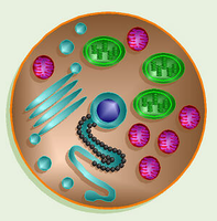
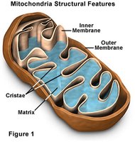
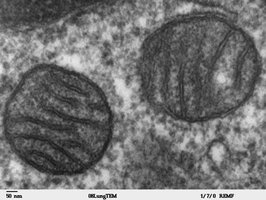
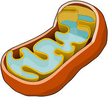
Mitochondria are surrounded by a double membrane. The inner membrane is highly folded (cristae) to increase surface area for reactions. The matrix is the internal space where metabolic processes occur.
Outer Membrane: Smooth boundary.
Inner Membrane: Folded into cristae.
Matrix: Contains enzymes for respiration.
Function
Mitochondria are known as the "powerhouse" of the cell because they are the site of aerobic respiration, producing energy (ATP) for cellular activities. The process requires oxygen:
Aerobic Respiration Equation:
Distribution in Cells
Cells with high metabolic rates (e.g., muscle cells, sperm cells) contain many mitochondria to meet energy demands. Inactive cells (e.g., fat cells) have fewer mitochondria.
Ribosomes
Structure and Function
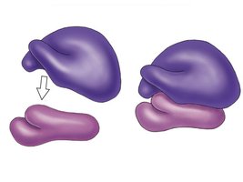
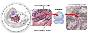
Ribosomes are made in the nucleolus and can be bound to the rough endoplasmic reticulum or float freely in the cytoplasm. They are responsible for protein synthesis.
Structure: Composed of a large and small subunit.
Function: Translate mRNA into proteins.
Endoplasmic Reticulum (ER)
Structure and Types
The ER is an interconnected network of membrane-enclosed sacs connected to the nuclear envelope and extending throughout the cell. There are two types:
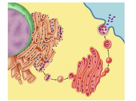
Rough ER: Ribosomes on surface; synthesizes proteins for secretion.
Smooth ER: No ribosomes; synthesizes lipids and detoxifies chemicals.
Vesicles
Structure and Function
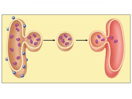
Vesicles are small, membrane-bound sacs used to transport materials within the cell. They bud from the ER and fuse with the Golgi apparatus or cell membrane.
Function: Transport proteins, waste, and other substances.
Golgi Apparatus
Structure and Function
The Golgi apparatus processes and modifies proteins from the rough ER, repackaging them in vesicles for secretion outside the cell.
Lysosomes
Structure and Function
Lysosomes are generally spherical with a single membrane, formed from Golgi vesicles. They contain digestive enzymes for breaking down ingested food, damaged organelles, or the cell itself.
Function: Intracellular digestion and recycling.
Plant Cell Organelles
Unique Features
Plant cells contain organelles not found in animal cells, including chloroplasts, a large central vacuole, and a cell wall.
Chloroplast: Site of photosynthesis; contains chlorophyll.
Vacuole: Maintains turgor pressure and stores substances.
Cell Wall: Rigid, structural layer made of cellulose.
Chloroplasts
Structure: Double membrane; contains chlorophyll.
Function: Site of photosynthesis, producing glucose.
Vacuole
Structure: Fluid-filled sac surrounded by tonoplast.
Function: Storage of food, water, waste; maintains turgor pressure.
Cell Wall
Structure: Rigid layer made of cellulose.
Function: Maintains cell shape, prevents damage from excess water intake, freely permeable to water and molecules.
Summary Table: Comparison of Animal and Plant Cells
Feature | Animal Cell | Plant Cell |
|---|---|---|
Cell Wall | Absent | Present (cellulose) |
Chloroplasts | Absent | Present |
Vacuole | Small, temporary | Large, permanent |
Shape | Round/irregular | Rectangular/regular |
Energy Source | Food (glucose) | Sunlight (photosynthesis) |
Additional info: These notes expand on the original content by providing academic context, definitions, and examples for each organelle and cell structure, ensuring completeness and clarity for exam preparation.
