 Back
Topic2_Biol186_W2026 (2).pdf
Study Guide - Smart Notes
 Tailored notes based on your materials, expanded with key definitions, examples, and context.
Introduction to Cell Biology
Cell biology is a foundational discipline in biology, focusing on the structure, function, and processes of cells—the basic units of life. Understanding cells is essential for exploring physiology, genetics, and molecular biology.
The Cell Theory
Historical Context and Principles
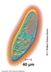
All living organisms are composed of cells, which may be unicellular or multicellular.
Cells are the basic units of structure and function in living things.
All cells arise from pre-existing cells through cell division.
Units of Life
Unicellular and Multicellular Organisms
Unicellular organisms consist of a single cell (e.g., bacteria, archaea, some protists).
Multicellular organisms are composed of many cells organized into tissues and organs (e.g., plants, animals, fungi).
Cells vary in size, shape, and function, but all share certain fundamental features.
Studying Cells
Microscopy and Cell Visualization
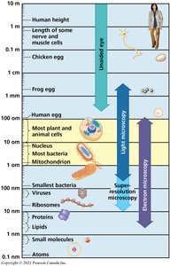
Microscopy is essential for studying cells, as most cells are too small to be seen with the naked eye.
Light microscopy allows visualization of most plant and animal cells, nuclei, and some organelles.
Electron microscopy provides higher resolution, enabling observation of smaller structures such as viruses, ribosomes, and membranes.
Magnification is the increase in apparent size, while resolution is the ability to distinguish two points as separate.
Types of Microscopy
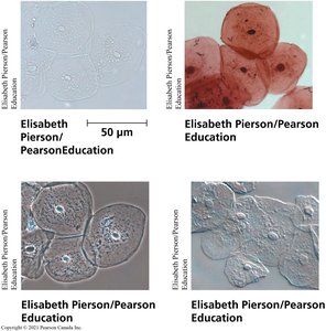
Brightfield microscopy: Standard light microscopy, can be used with or without stains.
Phase contrast and differential interference contrast microscopy: Enhance contrast in unstained cells.
Scanning Electron Microscopy (SEM): Provides detailed surface images.
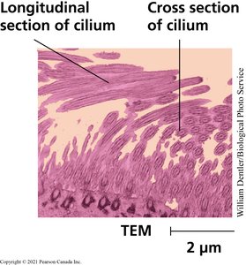
Transmission Electron Microscopy (TEM): Reveals internal cell structures.

Cell Fractionation
Cell fractionation is a technique used to isolate specific organelles from cells for further study. This is achieved by homogenizing cells and using differential centrifugation to separate components based on size and density.
Homogenization: Breaking cells open to release organelles.
Differential centrifugation: Sequentially increasing centrifugal force to pellet cellular components.

Prokaryotes
Structure and Features
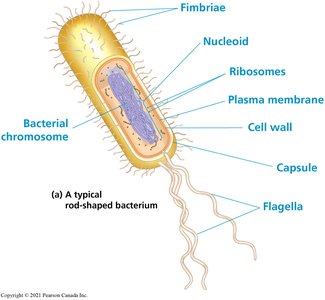
Prokaryotes include bacteria and archaea. They lack membrane-bound organelles and a nucleus, but possess all the essential components for life.
Key components: Plasma membrane, cytosol, DNA (nucleoid), ribosomes.
Additional structures: Cell wall, capsule, fimbriae, flagella.
Prokaryotic cells are typically 1–5 µm in size, but some can be much larger.
Prokaryotic Diversity and Size
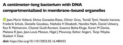
Most bacteria are small, but some, such as Ca. Thiomargarita magnifica, can reach centimeter lengths.
Prokaryotes exhibit a wide range of morphologies and ecological roles.
From Prokaryotes to Eukaryotes
Surface Area-to-Volume Ratio
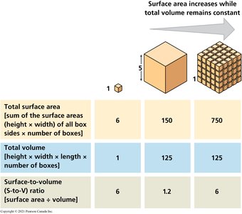
As cells increase in size, their volume grows faster than their surface area, limiting the rate of material exchange with the environment. This principle explains why most cells are small and why multicellular organisms are composed of many cells rather than a few large ones.
Single Cube | 5x5x1 Cubes | 5x5x5 Cubes | |
|---|---|---|---|
Total Surface Area | 6 | 150 | 750 |
Total Volume | 1 | 125 | 125 |
Surface-to-Volume Ratio | 6 | 1.2 | 6 |
Cell Membranes
Structure and Function
The plasma membrane is a selectively permeable barrier composed of a phospholipid bilayer with embedded proteins. It regulates the movement of substances into and out of the cell and facilitates communication with the environment.
Phospholipid bilayer: Hydrophilic heads face outward, hydrophobic tails inward.
Proteins: Serve as channels, receptors, enzymes, and structural components.
Carbohydrate side chains: Involved in cell recognition and signaling.

Eukaryotes: Animal Cell Structure
Key Organelles and Their Functions
Nucleus: Contains genetic material (DNA) and controls cellular activities.
Endoplasmic Reticulum (ER): Rough ER synthesizes proteins; smooth ER synthesizes lipids and detoxifies chemicals.
Golgi Apparatus: Modifies, sorts, and packages proteins and lipids for transport.
Mitochondria: Site of cellular respiration and ATP production.
Lysosomes: Contain hydrolytic enzymes for digestion of macromolecules.
Peroxisomes: Break down fatty acids and detoxify harmful substances.
Cytoskeleton: Provides structural support and facilitates cell movement.

Summary Table: Prokaryotic vs. Eukaryotic Cells
Feature | Prokaryotes | Eukaryotes |
|---|---|---|
Nucleus | No | Yes |
Membrane-bound organelles | No | Yes |
Size | 1–5 µm | 10–100 µm |
Examples | Bacteria, Archaea | Plants, Animals, Fungi, Protists |
Conclusion
Cells are the fundamental units of life, exhibiting remarkable diversity in structure and function. Understanding their organization, the differences between prokaryotic and eukaryotic cells, and the principles governing their size and complexity is essential for further study in biology.
