 Back
BackCell Structure and Function: The Nucleus, Ribosomes, and Endomembrane System
Study Guide - Smart Notes
 Tailored notes based on your materials, expanded with key definitions, examples, and context.
The Nucleus and Its Components
Nuclear Envelope and Structure
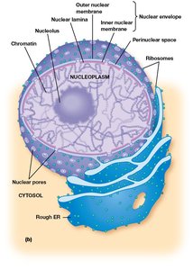
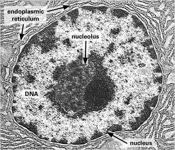
The nucleus is the control center of the cell, responsible for storing genetic information and coordinating cellular activities. It is surrounded by a double membrane called the nuclear envelope, which separates the nucleoplasm from the cytosol. The envelope consists of an inner and outer membrane, with the outer membrane continuous with the endoplasmic reticulum (ER).
Nuclear envelope: Double membrane structure enclosing the nucleus.
Perinuclear space: The space between the inner and outer nuclear membranes.
Nuclear pores: Openings in the envelope that regulate transport of molecules in and out of the nucleus.
Nucleoplasm: The semi-fluid matrix inside the nucleus.
Chromatin: DNA and associated proteins found within the nucleus.
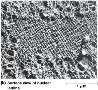
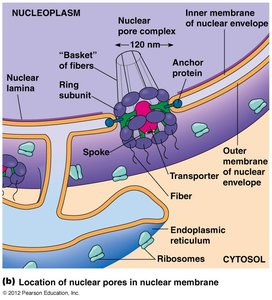
Nuclear Lamina
The nuclear lamina is a network of intermediate filaments located just beneath the inner nuclear membrane. It provides structural support, maintains nuclear shape, and organizes chromatin.
Intermediate filaments: Cytoskeletal components forming the lamina.
Scaffolding: Supports the nuclear envelope and anchors nuclear pores.
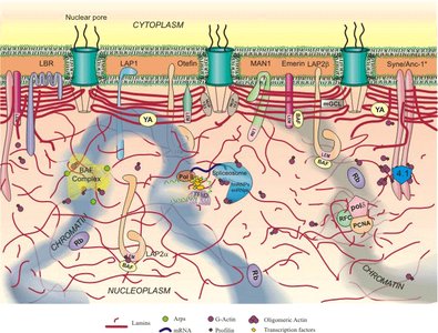
Nuclear Pores and Transport
Nuclear pores are large protein complexes embedded in the nuclear envelope, forming channels for regulated exchange of molecules between the nucleus and cytoplasm. The nuclear pore complex (NPC) controls movement of proteins, RNAs, and other macromolecules.
NPC: Nuclear pore complex, a multi-protein structure regulating transport.
Transported molecules: mRNA, ribosomal subunits, proteins, and regulatory factors.
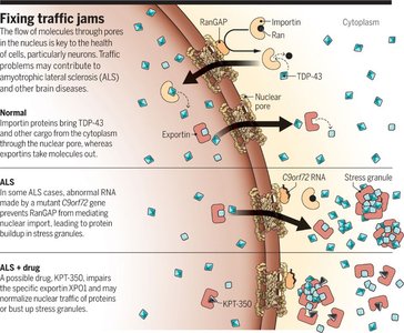
Nuclear Transport and Disease
Disruptions in nuclear transport are linked to neurodegenerative diseases, such as amyotrophic lateral sclerosis (ALS). Abnormal RNA or protein accumulation can block nuclear pores, impairing cellular function.
Importin/Exportin: Proteins mediating nuclear import and export.
ALS: Disease associated with defective nuclear transport.
Nucleolus
Structure and Function
The nucleolus is a dense region within the nucleus, responsible for ribosomal RNA (rRNA) synthesis and ribosome assembly. Its size correlates with the rate of protein synthesis in the cell.
rRNA genes: Clustered in the nucleolus.
Protein synthesis: The process of translation, where ribosomes produce proteins.
Ribosomes
Structure and Types
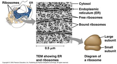
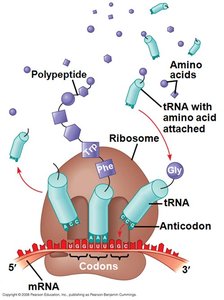
Ribosomes are ribonucleoprotein complexes found in all cell types. They are not membrane-bound and are responsible for protein synthesis. Ribosomes consist of two subunits, which differ between prokaryotes and eukaryotes.
Prokaryotic ribosome: 50S (large) + 30S (small) = 70S assembled
Eukaryotic ribosome: 60S (large) + 40S (small) = 80S assembled
Free ribosomes: Located in cytosol, synthesize proteins for internal use.
Bound ribosomes: Attached to ER, synthesize proteins for secretion or membrane insertion.
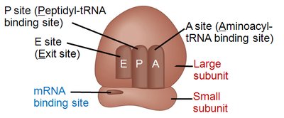
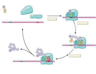
Ribosome Function: Translation
Translation is the process by which ribosomes synthesize proteins from mRNA templates. It involves three main stages: initiation, elongation, and termination.
Initiation: Ribosome assembles on mRNA, tRNA binds to start codon (AUG).
Elongation: tRNAs bring amino acids, which are added to the growing polypeptide chain.
Termination: Stop codon is recognized, and the completed polypeptide is released.
Antibiotics and Ribosome Inhibition
Certain antibiotics inhibit prokaryotic ribosome function, blocking protein synthesis and serving as effective treatments for bacterial infections.
Spectinomycin: Interferes with mRNA interaction with 30S ribosome.
Tetracycline: Binds irreversibly to 30S subunit, prevents tRNA entry into A site.
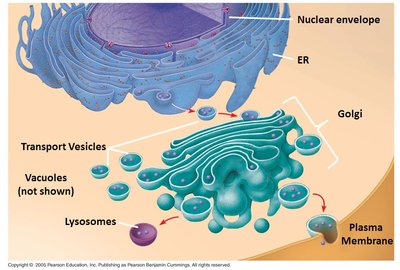
Endomembrane System
Components and Connections
The endomembrane system is a network of membranes and internal spaces in eukaryotic cells, including the nuclear envelope, ER, Golgi apparatus, lysosomes, vacuoles, and transport vesicles. These components are interconnected directly or via vesicle transport.
Nuclear envelope
Endoplasmic reticulum (ER)
Golgi apparatus
Lysosomes
Vacuoles
Transport vesicles
Plasma membrane
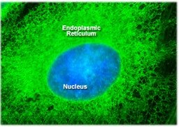
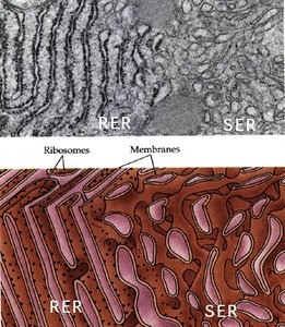
Endoplasmic Reticulum (ER)
The ER is a major organelle in eukaryotic cells, accounting for about half of the cell's membranes. It consists of two distinct regions: rough ER (with ribosomes) and smooth ER (without ribosomes).
Rough ER: Synthesizes proteins for secretion and membrane insertion.
Smooth ER: Synthesizes lipids, metabolizes carbohydrates, stores calcium, and detoxifies poisons.
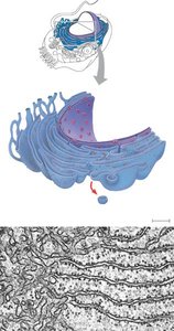
Golgi Apparatus
The Golgi apparatus is a system of flattened membranous sacs that receives transport vesicles from the ER. It modifies, sorts, and packages proteins and other molecules for transport to their final destinations.
Cis Golgi network (CGN): Entry side, receives vesicles from ER.
Trans Golgi network (TGN): Exit side, sorts and ships molecules.
Modification: Glycosylation of proteins, manufacture of polysaccharides.
Lysosomes
Lysosomes are membranous sacs containing hydrolase enzymes, which catalyze the hydrolysis of macromolecules. They function as digestive compartments, breaking down ingested material and cellular debris.
Hydrolases: Enzymes that add water to bonds, causing them to break.
Intracellular digestion: Breakdown of particles and organelles.
Phagocytosis: Ingestion of large particles, formation of phagosomes.
Autophagy: Degradation of old or damaged cell structures.
Vacuoles
Vacuoles are diverse maintenance compartments found in many cells. In plants, the central vacuole maintains fluid balance and turgor pressure. Other types include food vacuoles (phagosomes) and contractile vacuoles for osmoregulation.
Central vacuole: Stores water and organic compounds, maintains turgor pressure.
Food vacuole: Formed by phagocytosis, digests ingested material.
Contractile vacuole: Regulates water balance in protists.
Energy-Converting Organelles
Mitochondria and Chloroplasts
Mitochondria are the sites of cellular respiration, converting chemical energy from food into ATP. Chloroplasts are found only in photosynthetic eukaryotes and are the sites of photosynthesis, converting light energy into chemical energy.
Mitochondria: Present in most eukaryotes, including animals and fungi.
Chloroplasts: Present in plants and algae.
Endosymbiotic Theory
The endosymbiotic theory proposes that mitochondria and chloroplasts originated from prokaryotic cells engulfed by ancestral eukaryotes. Mitochondria evolved from proteobacteria, and chloroplasts from cyanobacteria.
Evidence: Similar size to prokaryotes, double membranes, prokaryotic ribosomes, circular genomes, sequence similarity to bacteria.
Gene transfer: Many organelle genes are now encoded in the nucleus.
Peroxisomes
Structure and Function
Peroxisomes are small, membrane-bound organelles similar in shape and size to lysosomes, but not part of the endomembrane system. They compartmentalize oxidative reactions and detoxify harmful substances.
Oxidative reactions: Breakdown of organic molecules, generating hydrogen peroxide (H2O2).
Detoxification: Catalase and peroxidase enzymes degrade H2O2 to water and oxygen.
Fatty acid breakdown: β-oxidation pathway.
Cytoskeleton
Components and Functions
The cytoskeleton is a network of protein filaments that organizes subcellular structures, provides mechanical support, and enables cell movement.
Microtubules: Spatial organization, intracellular transport.
Microfilaments: Cell shape, locomotion, muscle contraction.
Intermediate filaments: Mechanical strength, nuclear shape.
Cell Locomotion and Phagocytosis
Cell movement, such as neutrophil chasing bacteria, is driven by cytoskeletal motor proteins. Phagocytosis involves the formation of phagosomes and fusion with lysosomes for digestion.
Defective phagocytosis: Failure of phagocytic vesicle and lysosome fusion can result in persistent infection.
