 Back
Cell Structure and Membrane Function: Study Notes for General Biology
Study Guide - Smart Notes
 Tailored notes based on your materials, expanded with key definitions, examples, and context.
Cell Structure and Function
Animal Cells vs. Plant Cells
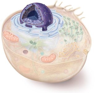
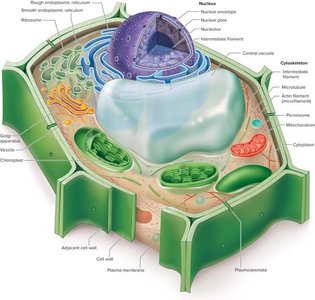
Animal and plant cells share many organelles, but also possess unique structures that distinguish them. Understanding these differences is fundamental to cell biology.
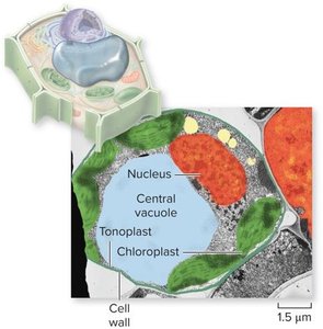
Plant Cells: Contain a cell wall, chloroplasts, and a large central vacuole.
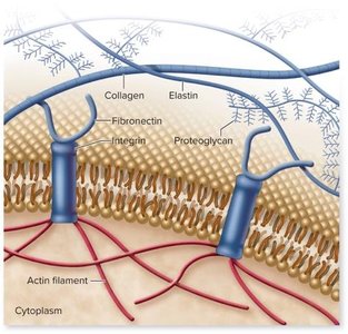
Animal Cells: Lack a cell wall and chloroplasts, but possess an extracellular matrix and centrioles.
Common Organelles: Both have nuclei, mitochondria, endoplasmic reticulum, Golgi apparatus, and ribosomes.
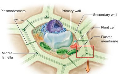
Cell Wall: Provides structural support and protection in plants; composed mainly of cellulose.
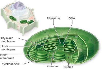
Chloroplasts: Site of photosynthesis in plants, containing thylakoid membranes and stroma.
Vacuole: Stores water, nutrients, and supports cell wall in plants.
Extracellular Matrix (ECM): In animals, a network of proteins (collagen, elastin, fibronectin) that influences cell behavior.
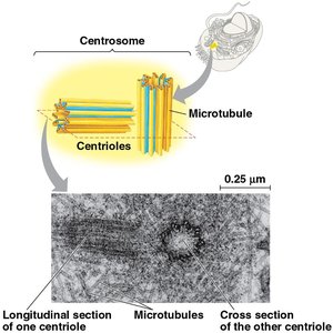
Centrioles: Organize microtubules during mitosis in animal cells.
Example: Red blood cells (animal) lack a cell wall, while plant cells have a rigid cell wall.
The Cytoskeleton
Components and Functions
The cytoskeleton is a dynamic network of protein filaments that provides structural support, facilitates cell movement, and organizes organelles within the cell.
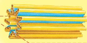
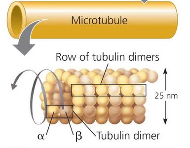
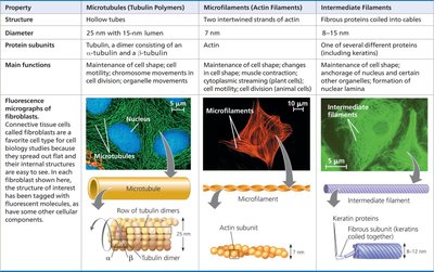
Microtubules: Hollow tubes made of tubulin dimers; involved in cell shape, organelle movement, chromosome separation, and controlling cilia/flagella.
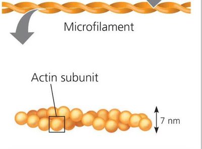
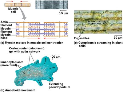
Microfilaments: Twisted double chains of actin; support cell shape, enable muscle contraction, and facilitate cellular motility (e.g., pseudopodia).
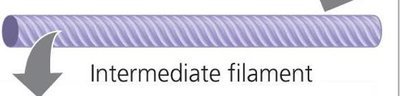
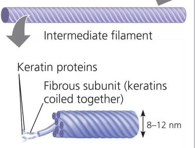
Intermediate Filaments: Fibrous proteins (e.g., keratin) coiled into cables; provide permanent structural support and anchor organelles.
Centrosomes and Centrioles: Microtubules grow out from centrosomes; centrioles are pairs of microtubule triplets in animal cells.
Example: Amoeboid movement is driven by microfilaments extending pseudopodia.


Comparison Table: Cytoskeletal Elements
This table summarizes the structure, protein subunits, and main functions of the three cytoskeletal components.
Property | Microtubules (Tubulin Polymers) | Microfilaments (Actin Filaments) | Intermediate Filaments |
|---|---|---|---|
Structure | Hollow tubes | Two intertwined strands of actin | Fibrous proteins coiled into cables |
Diameter | 25 nm with 15-nm lumen | 7 nm | 8–12 nm |
Protein Subunits | Tubulin (α and β dimers) | Actin | Keratin and other proteins |
Main Functions | Cell shape, motility, chromosome movement, organelle movement | Cell shape, muscle contraction, cytoplasmic streaming, cell motility, cell division | Cell shape, anchorage of nucleus/organelles, nuclear lamina formation |
Membrane Structure and Function
Fluid Mosaic Model
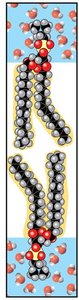
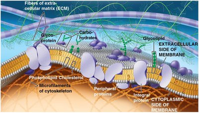
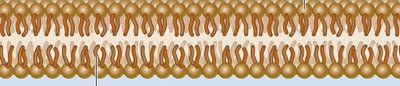
Cellular membranes are composed of a fluid mosaic of lipids, proteins, and carbohydrates. The main structural component is the phospholipid bilayer, which is amphipathic—containing both hydrophilic and hydrophobic regions.
Phospholipids: Form a bilayer with hydrophilic heads facing outward and hydrophobic tails inward.
Proteins: Integral and peripheral proteins serve as channels, carriers, and receptors.
Carbohydrates: Attached to proteins and lipids, functioning in cell recognition.
Example: The plasma membrane regulates entry and exit of substances.
Selective Permeability
Membrane structure results in selective permeability, allowing some substances to cross more easily than others. The permeability depends on the polarity and size of molecules.
Hydrophobic (nonpolar) molecules: Pass rapidly through the lipid bilayer (e.g., O2, CO2).
Hydrophilic (polar) molecules: Pass slowly or require transport proteins (e.g., sugars, ions).
Example: Water and glucose require channel or carrier proteins to cross the membrane.
Transport Proteins
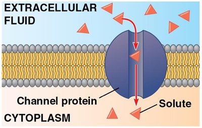
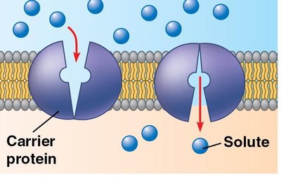
Transport proteins facilitate the movement of hydrophilic substances across the membrane. Channel proteins create hydrophilic tunnels, while carrier proteins bind and shuttle molecules.
Channel Proteins: Allow specific ions or molecules to pass through.
Carrier Proteins: Change shape to move substances across the membrane.
Example: Aquaporins (water channels) and GLUTs (glucose transporters).
Passive Transport
Diffusion
Diffusion is the passive movement of particles from regions of higher concentration to lower concentration, driven by the concentration gradient. No energy is required.
Dynamic Equilibrium: Occurs when molecules move equally in both directions across the membrane.
Concentration Gradient: The difference in concentration across a space.
Example: Oxygen diffuses into cells where its concentration is lower.
Osmosis
Osmosis is the diffusion of water across a selectively permeable membrane. Water moves from areas of lower solute concentration to higher solute concentration until equilibrium is reached.
Solute: Substance dissolved in a solvent.
Solvent: The medium that dissolves the solute (usually water).
Example: Water moves into plant cells, causing turgor pressure.
Tonicity
Tonicity describes the effect of a solution on cell volume, based on solute concentration.
Isotonic: Equal solute concentration inside and outside the cell; no net water movement.
Hypertonic: Higher solute concentration outside; cell loses water and shrinks (crenation in animals, plasmolysis in plants).
Hypotonic: Lower solute concentration outside; cell gains water and may swell.
Example: Red blood cells shrink in hypertonic solutions.
Facilitated Diffusion
Protein-Aided Transport
Facilitated diffusion is a form of passive transport where hydrophilic substances move across membranes via transport proteins. No energy is required.
Channel Proteins: Provide passageways for ions and small molecules.
Carrier Proteins: Undergo conformational changes to move substances.
Gated Channels: Open or close in response to stimuli (e.g., electrical signals in nerve cells).
Example: Potassium ion channels open in response to neurotransmitters.
Active Transport
Energy-Dependent Transport
Active transport moves solutes against their concentration gradients using energy, typically from ATP hydrolysis. All proteins involved are carrier proteins.
Sodium-Potassium Pump: Maintains membrane potential by pumping Na+ out and K+ into the cell.
Electrochemical Gradient: Combination of chemical and electrical forces driving ion movement.
Cotransport: Active transport of one solute indirectly drives transport of another.
Example: Plant cells use proton pumps and cotransporters to load sucrose into veins.
Equation: ATP hydrolysis provides energy for active transport:
Bulk Transport
Exocytosis and Endocytosis
Bulk transport moves large molecules across the membrane via vesicles, requiring energy.
Exocytosis: Vesicles fuse with the membrane to release contents outside the cell (e.g., insulin secretion).
Endocytosis: Cell takes in substances via vesicles; includes phagocytosis (solids) and pinocytosis (fluids).
Receptor-Mediated Endocytosis: Specific molecules are taken in after binding to cell surface receptors.
Example: Plant cells release cell wall material by exocytosis.
Additional info:
These notes cover key concepts from Chapters 6 and 7: cell structure, cytoskeleton, and membrane function, including transport mechanisms.
For further study, review the structure of matter and chemical elements (Chapter 2).
